
Image
|
Figure Caption
Fig. 2
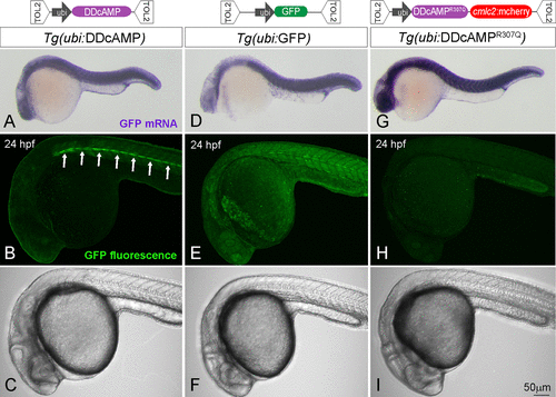
Figure 2. (top) Diagram of the TOL2 plasmids containing N41-GFP, N41R307Q-GFP, and GFP DNA under control of ubi promoter. (bottom) In situ hybridization for GFP mRNA in Tg(ubi:DDcAMP) (A), Tg(ubi:GFP) (D) and Tg(ubi:DDcAMPR307Q) (G) embryos at 24 hpf. Confocal acquisition of GFP signal in Tg(ubi:DDcAMP) (B, C), Tg(ubi:GFP) (E, F), and Tg(ubi:DDcAMPR307Q) (H, I) embryos at 24 hpf. Horizontal myoseptum is indicated by white arrows in panel B.
Figure Data
Acknowledgments
This image is the copyrighted work of the attributed author or publisher, and
ZFIN has permission only to display this image to its users.
Additional permissions should be obtained from the applicable author or publisher of the image.
Full text @ ACS Chem. Biol.