
Image
|
Figure Caption
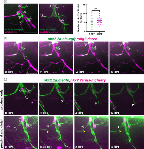
Fig. 2
Perineurial glia do not proliferate following spinal motor nerve injury. (a) (Left) Representative images of perineurial glial nuclei (open arrows) imaged with either motor axons (magenta, left) or perineurial glial membrane (green, right) at 5 dpf. (Right) Quantification of the number of nkx2.2a+ nuclei on a single spinal motor nerve in 5 (green, n = 10 nerves in 5 larvae, mean: 4.8 ± 0.79 nuclei) and 6 dpf (magenta, n = 17 nerves in 5 larvae, mean: 6.1 ± 0.44 nuclei) larvae (p = .1272). (b) Representative images from a time-lapse movie of nkx2.2a+ nuclei (green) in an injured 6 dpf larva. The dashed circle indicates the injury site (n = 4 nerves in 3 larvae). (c) Representative still images from a time-lapse movie of nkx2.2a+ nuclei (magenta, open arrowheads) in injured 5 to 6 dpf larvae exhibiting either proximal only bridging (top panels, n = 6 nerves in 4 larvae) or proximal and distal bridging (bottom panels, n = 4 nerves in 3 larvae). Solid white arrows follow the proximal end and yellow arrows follow the distal end of the perineurial glial bridge (green). Asterisks indicate nkx2.2a+ cells that are not perineurial glial nuclei. Scale bars, 25 μm.
Acknowledgments
This image is the copyrighted work of the attributed author or publisher, and
ZFIN has permission only to display this image to its users.
Additional permissions should be obtained from the applicable author or publisher of the image.
Full text @ Glia