
Image
|
Figure Caption
Fig. 5
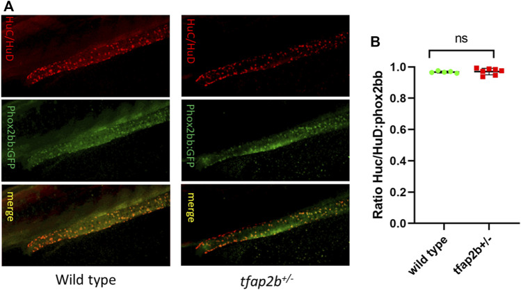
Differentiated enteric neurons in tfap2b +/? fish. (A) Confocal microscope images of HuC/HuD and phox2bb:GFP positive cells in wild type and tfap2b +/? F2 fish. (B) No significant difference in the ratio of HuC/HuD and phox2bb:GFP positive cells in wild type and tfap2b +/? F2 fish was found (p > 0.05, unpaired t-test).
Figure Data
Acknowledgments
This image is the copyrighted work of the attributed author or publisher, and
ZFIN has permission only to display this image to its users.
Additional permissions should be obtained from the applicable author or publisher of the image.
Full text @ Front Cell Dev Biol