Image
Figure Caption
Fig. 5
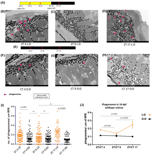
Zebrafish outer segment phagocytosis (OSP) is regulated by external light sources. (A,E) Time course highlighting sample collection time (red x), and light-dark cycle. Samples were collected at ZT 4, ZT 9, and ZT 17 from 16 days post fertilization (dpf) wild-type zebrafish retinae. At each time point, one cohort had undergone a regular 14:10 light:dark cycle. The other had been raised in dark from ZT 10 the day prior to collection and were euthanized under red light. (B?D, G,H) Representative TEM images of wild-type retinae at 16 dpf, with phagosomes indicated (pink arrows). Scale bar; 5 ?m. (I) Scatter plots of phagosome/?m of retinal pigment epithelium (RPE) in dark-reared and control retinas at ZT 4 and ZT 17 (OSP peaks) and ZT 9 (OSP trough). Fish raised in the dark for have ~35%?55% reductions in the number of RPE phagosomes containing engulfed OS material in comparison with controls at ZT 4 and ZT 17 respectively. Statistical analysis carried out by student's t test between individual groups. RPE of >570 ?m was measured per animal. N = 3 animals (6 retinae) per group per time point. (J) Trend line demonstrating the level of RPE phagosomes present in the dark-raised wild-type eyes (black) compared with controls exposed to the regular light-dark cycle (orange) at defined time points throughout the day. A two-way anova, followed by Turkey's multiple comparison testing revealed no significant difference between CT 4 CT 9 or CT 17 (black), but a significant difference between ZT 4 versus ZT 9 (p = .0229), ZT 9 versus ZT 17 (p ? .0001), and ZT 4 versus ZT 17 (p = .003) (orange)
Acknowledgments
This image is the copyrighted work of the attributed author or publisher, and
ZFIN has permission only to display this image to its users.
Additional permissions should be obtained from the applicable author or publisher of the image.
Full text @ FASEB J.

