
|
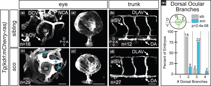
Fig. 5.
Ocular vasculature but not trunk vasculature is disrupted in shutdown corner. Ocular a, c) and trunk b, d) vasculature at 48 hpf [grayscale, Tg(kdrl:mCherry-ras)]. 3D renderings, lateral views. a′, c′) Hyaloid network; superficial vasculature cropped away in FluoRender. c) Arrowheads indicate ectopic branches; asterisk indicates a morphologically abnormal vessel. e) Quantification of the number of superficial ocular vessels in the dorsal half of the eye at 48 hpf. Dashed line in schematic demarcates the dorsal (D) and ventral (V) halves of the eye. Sample size (n) in images. a–d) Arrows pair labels with corresponding vessels. le, lens; DCV, dorsal ciliary vein; NCA, nasal ciliary artery; OV, optic vein; DLAV, dorsal longitudinal anastomosing vessel; aISV, arterial intersegmental vessel; A, anterior; P, posterior; D, dorsal; V, ventral. Scale bar, 50 µm.