
|
Fig. 1.
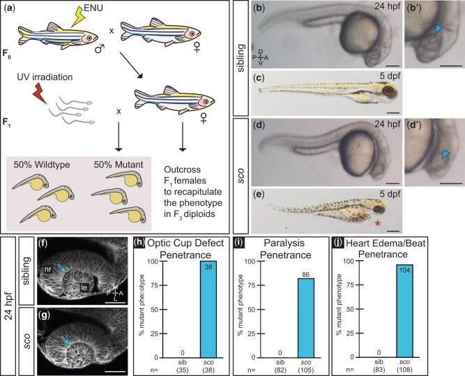
Shutdown corner, isolated in a haploid screen, exhibits a gross morphological defect of the optic cup. a) Haploid mutagenesis screen strategy. b, d) A defect in optic cup morphology is visible at 24 hpf. Lateral view of sibling b) and sco mutant d) diploid embryos under dissecting stereomicroscope. Zoomed views of sibling b?) and sco d?) lens regions (arrowheads), in which the lens is difficult to discern in the sco mutant. c, e) sco mutants exhibit heart edema (asterisk) and die around 5 dpf. f, g) Optic cup morphogenesis in live-imaged, membrane-labeled samples: a lens forms in sibling f) and sco mutants g) and is enwrapped by the developing retina in both. Dorsal view, single confocal section of 24 hpf Tg(bactin2:EGFP-CAAX) embryos. Arrows indicate the separation f) or apparent close association g) between the lens and neural retina. The sco mutant 24 hpf optic cup h), 3 dpf paralysis i), and 3 dpf heart edema/slowed heartbeat j) phenotypes are highly penetrant when screened on confocal or stereomicroscope. (h: sib = 0%, sco = 100%; i: sib = 0%, sco = 81.9%; j: sib = 0%, sco = 96.3%). A, anterior; L, lateral; le, lens; M, medial; nr, neural retina; P, posterior. Scale bar: b, b?, d, d?: 170 �m; c, e: 310 �m; and f, g: 50 �m.