
|
Fig. 6
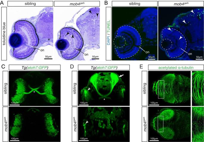
(A) At 3 dpf, toluidine blue-stained sections displayed pyknotic nuclei (arrowhead) dispersed throughout the retina and tectum of mob4geh homozygotes, not siblings (n = 4 per genotype). (B) Abundant apoptosis within the retina and tectum of 3-dpf-old mob4geh homozygotes was detected by TUNEL assay (n = 8 per genotype). (C) In representative ventral views (Z-stack), the optic chiasm (asterisk) was highlighted by Tg(ath7:GFP) within 3-dpf-old siblings but not mob4geh homozygotes (n = 3 per genotype). (D) In representative Z-stacks, axons of retinal ganglion cell marked by Tg(ath7:GFP) project contralaterally via the optic chiasm (asterisk) from the retina (arrowhead) onto the tectum (arrow) of 3-dpf-old siblings (n = 4). In contrast, axons were not formed by retinal ganglion cells (arrowhead) of mob4geh homozygotes (n = 4). (E) In Z-stacks of 3-dpf-old larvae, antibodies against acetylated ?-tubulin revealed defective neurite formation within the tectum of mob4geh homozygotes. Boxed areas are shown in higher magnification (n = 3 per genotype). Scale bar sizes are indicated.