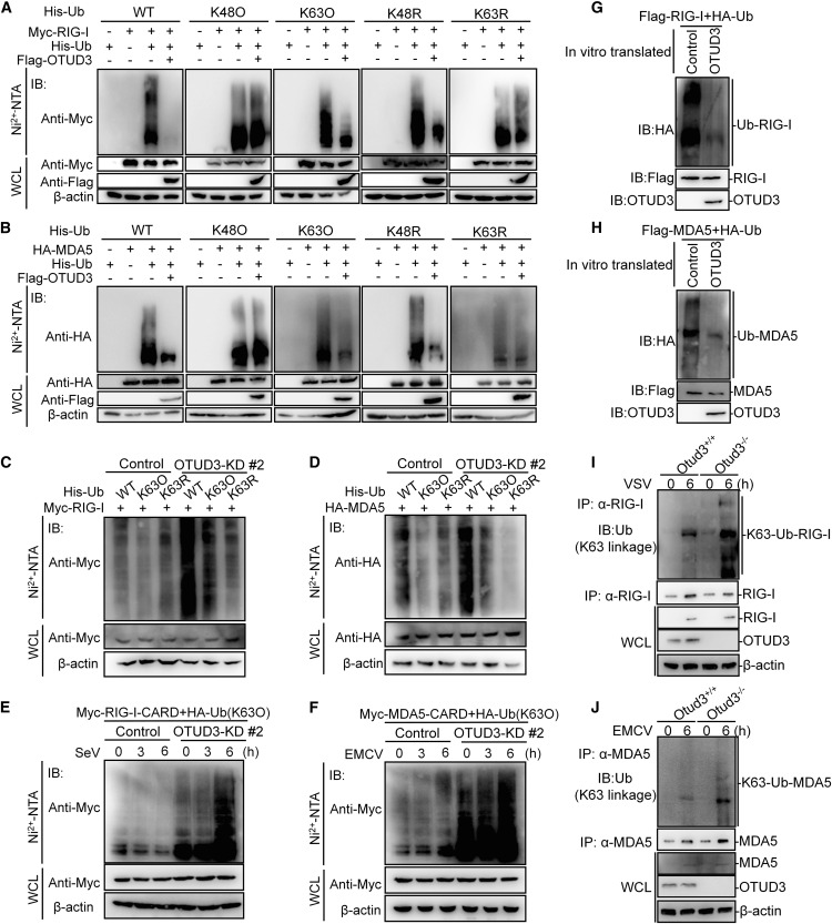
Fig. 2 Figure 2. OTUD3 inhibits RLR signaling by removing K63-linked ubiquitin chain on RIG-I and MDA5 (A and B) Immunoblotting (IB) for polyubiquitination of whole-cell lysates (bottom) and affinity purification with Ni2+-NTA resin (top) from HEK293T cells transfected with Myc-RIG-I (A) or HA-MDA5 (B) and His-tagged wild-type(WT) ubiquitin, His-tagged K48-only ubiquitin (K48O) (in which all lysine residues are mutated to arginine residues except lysine 48), His-tagged K63-only ubiquitin (K63O) (in which all lysine residues are mutated to arginine residues except lysine 63), His-tagged K48R ubiquitin (K48R) (in which only lysine 48 is mutated to arginine), or His-tagged K63R ubiquitin (K63R) (in which only lysine 48 is mutated to arginine), together with the empty vector or FLAG-OTUD3. (C and D) IB for polyubiquitination of whole-cell lysates and affinity purification with Ni2+-NTA resin from the control and OTUD3 knockdown (OTUD3-KD#2) HEK293T cells transfected with Myc-RIG-I (C) or HA-MDA5 (D) (3 ?g/each), together with His-WT ubiquitin, His-K63O ubiquitin, or His-K63R ubiquitin (4 ?g) for 24 h. (E and F) IB for polyubiquitination of whole-cell lysates and affinity purification with Ni2+-NTA resin from the control and OTUD3 knockdown (OTUD3-KD#2) HEK293T cells transfected with Myc-RIG-I-CARD (E) or Myc-MDA5-CARD (F) (3 ?g/each) and His-K63O ubiquitin (4 ?g) for 24 h, then infected with SeV or EMCV for the indicated times. (G and H) In vitro deubiquitination analysis of RIG-I (G) or MDA5 (H) by OTUD3. The lysates of HEK293T cells transfected with FLAG-RIG-I or FLAG-MDA5 (5 ?g) together HA-ubiquitin (5 ?g) were eluted with FLAG peptides and then incubated with the synthesized OTUD3 by an in vitro transcription and translation kit. The ubiquitination was detected by IB with anti-HA antibody. (I and J) IB for K63-linked polyubiquitination of endogenous RIG-I (I) or MDA5 (J) in WT and Otud3?/? BMDCs (1× 10⁸) stimulated with VSV or EMCV for 6 h. IP, immunoprecipitation; IB, immunoblotting; WCL, whole-cell lysate. Data are representative of three independent experiments (A?J).
Image
Figure Caption
Acknowledgments
This image is the copyrighted work of the attributed author or publisher, and
ZFIN has permission only to display this image to its users.
Additional permissions should be obtained from the applicable author or publisher of the image.
Full text @ Cell Rep.

