Fig. 7
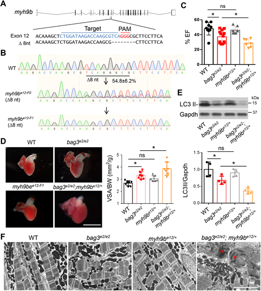
The stable myh9b heterozygous mutant deteriorated bag3 cardiomyopathy at adult stage. (A) Schematic of the predicted MMEJ-inducing myh9b genetic lesion of 8-nucleotides generated by injection of an sgRNA targeting sequences within the 12th exon. Dashed lines indicate an 8-nucleotide-long deletion. (B) Chromographs illustrating the sequences of the wild-type myh9b and the mutant alleles with the predicted 8-nucleotide-long deletion in F0 and F1 fish. (C) Quantification of cardiac function. Ejection fraction (EF) (in %) measured by using echocardiography in bag3e2/e2;myh9be12/+ double-mutant fish compared to single-mutant and WT control fish at 6 months; n=7-11, one-way ANOVA. (D) Left panel: Representative images of isolated hearts from fish as indicated. Right panel: Quantification of ventricular surface area (VSA) normalized to the body weight (BW) of fish at 6 months; n=7, one-way ANOVA. (E) Western blotting (top) and quantification LC3 II protein levels (bottom) of hearts from bag3e2/e2;myh9be12/+ double-mutant fish, bag3e2/e2, myh9be12/+ or WT control fish at 6 months. Levels of Gapdh were used as control. n=4, one-way ANOVA. (F) TEM images of bag3e2/e2;myh9be12/+ double-mutant fish hearts, bag3e2/e2, myh9be12/+ and WT controls at 6 months. Asterisks indicate Z-disc aggregation. Scale bar: 2 µM (D). *P<0.05. ns, not significant.

