
|
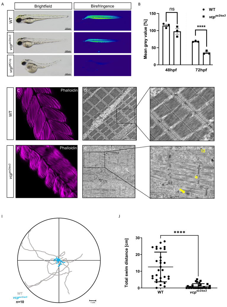
Fig. 2
Genetic loss of vcp leads to skeletal muscle dysfunctions. (A) Lateral view of brightfield and birefringence images for WT, vcpex3/ex3, and VCP-MO-injected embryos at 72 hpf. (B) Densitometric analysis of the birefringence signal at 72 hpf reveals significantly reduced signal intensity in vcpex3/ex3 embryos, implying impaired skeletal muscle organization, whereas birefringence signal intensity is unaltered at 48 hpf (48 hpf: N = 3, n = 5/8, p = 0.6690; 72 hpf: N = 3, n = 8/15; p = 0.0001; two-tailed t-test). Error bars indicate s.d.; **** p < 0.0001; ns, not significant. (C,F) Confocal microscopic analysis of phalloidin-stained skeletal muscles of WT and vcpex3/ex3 embryos at 72 hpf. vcpex3/ex3 embryos show disrupted muscle fibers compared to the clutchmates. (D,E,G,H) Electron microscopic pictures of WT and vcpex3/ex3 skeletal muscle embryos at 72 hpf. vcpex3/ex3 embryos show damaged mitochondria (M) and increased electron-dense inclusion bodies (?) in addition to the disrupted muscle fibers (*). (I) Touch-evoked assay reveals reduction in responsiveness upon mechanical stimulus in vcpex3/ex3 embryos, as shown in the motion trace diagram. (J) Quantification of the total swim distance shows significantly reduced motility in vcpex3/ex3 compared to WT embryos (N = 3, n = 10, mean � S.D; p < 0.0001; two-tailed t-test). Error bars indicate s.d.; **** p < 0.0001. ns: not significant.