Image
Figure Caption
Figure 5
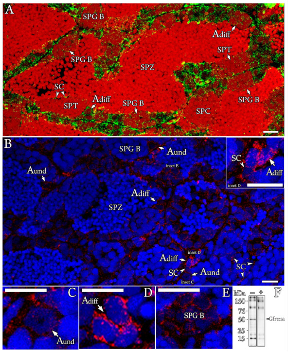
Figure 5. Cellular localization of Gfr?1a in zebrafish testis. (A?E) Immunofluorescence for Gfr?1a (green?A; red?B?E) in testis sections of sexually mature zebrafish. The spermatogonial generations, including type A undifferentiated spermatogonia (Aund), type A differentiated spermatogonia (Adiff) and type B spermatogonia (SPG B), were immunoreactive to Gfra1a, although staining patterns among them varied according to developmental stage. The signal was not found in spermatocytes (SPCs), spermatids (SPTs) and spermatozoa (SPZ). Note that Sertoli cells (SCs) contacting germ cells at different stages of development were also immunoreactive to Gfr?1a. Cell nuclei were counterstained with propidium iodide (A) or Hoechst (B?E). Scale bars: 15 Ám. (F) Gfr?1a (approximately 52 kDa (kilodaltons)) immunoblots of whole testes with (+) or without (?) preadsorbed antibodies, confirming the presence of the protein in the zebrafish testes and antibody specificity.
Figure Data
Acknowledgments
This image is the copyrighted work of the attributed author or publisher, and
ZFIN has permission only to display this image to its users.
Additional permissions should be obtained from the applicable author or publisher of the image.
Full text @ Cells

