
|
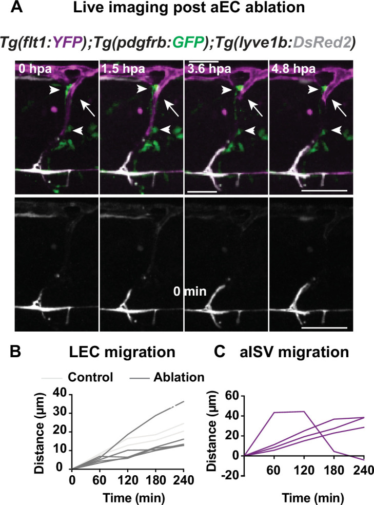
Figure 2—figure supplement 3. (A) Confocal stack images from time-lapse post multi-photon laser ablation in 2 days post fertilization (dpf) Tg(flt1:YFP) (magenta); TgBAC(pdgfrb:GFP) (green) and Tg(?5.2lyve1b:DsRed2) (grey) embryos. Arrowheads indicate remained GFP+ MCs without arterial intersegmental vessel (aISV). White arrows indicate the ablated site of aISV. Scale bar: 50 ?m (Figure 2?figure supplement 2); arterial endothelial cell (aEC) ablation with multi-photon laser. (A?B) Quantification of lymphatic endothelial cell (LEC) (n = 4) and aISV (n = 4) migration distance post two-photon laser ablation.