IMAGE

Fig. 3

ID
ZDB-IMAGE-220328-7
Source
Figures for Kuang et al., 2022
Image
Figure Caption

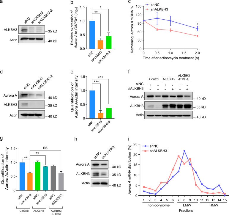
Fig. 3 Knockdown of ALKBH3 attenuates the stability and translation of <italic>Aurora A</italic> mRNA.

a Western blotting analysis of ALKBH3 in RPE-1 cells treated with the indicated siRNAs. Actin was used as a loading control. b Quantitative real-time PCR (qRT-PCR) analysis of Aurora A in RPE-1 cells transfected with the indicated siRNAs. GAPDH was served as an internal control. c Quantitative RT-PCR of Aurora A mRNA in ALKBH3-depleted and control cells treated with actinomycin D (5 μg/mL) at the indicated times. dg Western blotting of Aurora A in RPE-1 cells transfected with the siRNAs or plasmids as shown. Actin, a loading control. The intensity of Aurora A was measured based on the normalization to Actin. h Western blotting analysis of ALKBH3 and Aurora A in RPE-1 cells infected with lentivirus carrying shRNA targeting ALKBH3. Actin was used as a loading control. NC, negative control. i Polysome profiling of Aurora A mRNA in cells infected with lentivirus carrying shRNA targeting ALKBH3 after sucrose gradient centrifugation. Data are shown as the means ± SD from at least three independent experiments. Student’s t-test; ns, not significant; *P < 0.05, **P < 0.01, ***P < 0.001.

Acknowledgments
This image is the copyrighted work of the attributed author or publisher, and ZFIN has permission only to display this image to its users. Additional permissions should be obtained from the applicable author or publisher of the image. Full text @ Cell Discov