Fig 4
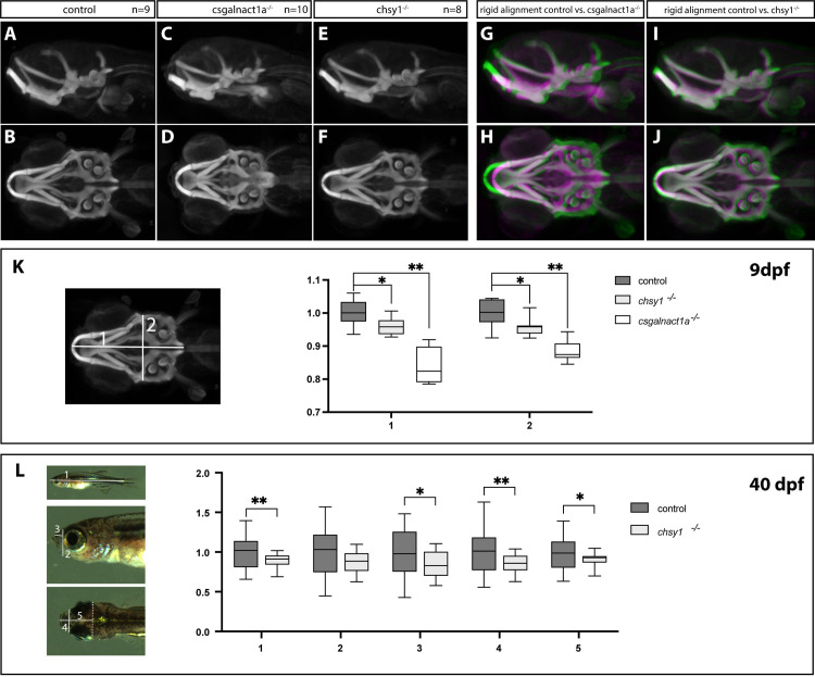
Maximum projections of average patterns generated from control, csgalnact1a-/- and chsy1-/- alcian blue stained larvae at 9 dpf show the pharyngeal cartilage structures in a ventral view (A-F). Maximum projections for each mutant (magenta) aligned to the control (green) are displayed using a rigid transformation (G-J). Measurements of the length and width of the head skeleton of 9 dpf larvae were performed on maximum projection images as shown in the image to the right (K) and plotted as a factor of control larvae (n = 8 for all genotype groups) (K). csgalnact1a-/- and chsy1-/- larval head skeleton is significantly smaller compared to control (K). Measurements of the standard body length (1) and different other measurements of the head (2?5) are indicated on images of 40 dpf old juvenile fish (L). chsy1-/- juveniles are significantly shorter and have a smaller head compared to control larvae (chsy1-/- n = 17, control n = 27) (L). Statistical significance is indicated by * for p-values <0.05 and ** for p-values <0.005.

