
Image
|
Figure Caption
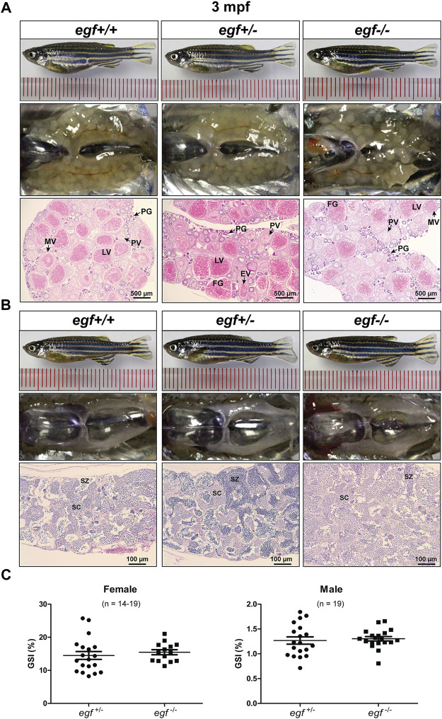
FIGURE 3 Gonadal development of egf mutant at 3 mpf. (A) Anatomical and histological examination of the ovary in egf mutant (egf?/?) and controls (egf+/+ and egf+/?). The egf-deficient follicles developed normally. (B) Anatomical and histological examination of the testis in egf mutant (egf?/?) and controls (egf+/+ and egf+/?). The spermiogenesis was normal in egf-deficient males. (C) GSI in females (n = 14?19) and males (n = 19). PG, primary growth; PV, previtellogenic; EV, early vitellogenic; MV, mid-vitellogenic; LV, late vitellogenic; FG, full-grown; SC, spermatocytes; SZ, spermatozoa.
Figure Data
Acknowledgments
This image is the copyrighted work of the attributed author or publisher, and
ZFIN has permission only to display this image to its users.
Additional permissions should be obtained from the applicable author or publisher of the image.
Full text @ Front Cell Dev Biol