
|
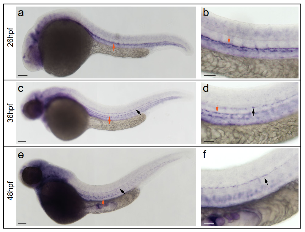
Fig. S4 In situ hybridization against vegfc using a milder proteinaseK treatment reveals expression at the horizontal myoseptum. a, b) vegfc transcripts were detected in the hypochord, dorsal aorta (arrow) and in developing intersegmental arteries at 26hpf. b) Higher magnification of the trunk region with prominent staining of the hypochord (arrow). c, d) At 36hpf vegfc transcripts were detected in cells at the HM (black arrow), the hypochord and the dorsal aorta (orange arrow). d) Higher magnification of the trunk region. e, f) Expression of vegfc within the DA and in cells at the HM persist at 48hpf. f) High magnification of the trunk focusing at the HM level. Scale bars in a, c, e: 100?m; b, d, f: 50?m. hpf: hours post fertilization, DA: dorsal aorta, HM: horizontal myoseptum.