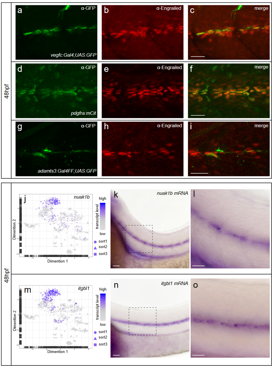
Fig. S11 Fibroblasts at the horizontal myoseptum express Engrailed proteins. a-i) HM region of 48hpf embryos that were stained with anti-Engrailed (red) and anti-GFP (green) antibodies. a-c) All cells expressing the vegfc:Gal4FF; UAS: GFP reporter at the HM do co-express Engrailed proteins. d-f) fpdg-fra:mCitrine positive fibroblasts at the HM are also positive for Engrailed. g-i) Partial z-projections of the HM region reveal a co-expression of the adamts3 reporter and Engrailed proteins within fibroblasts. j, m) Two additional examples of genes whose transcripts are highly enriched within fibroblast cluster 2 are nuak1b and tgbl1 (see Supplementary Tables 1-3). The mRNA of both genes (k, l, n, o) can be detected specifically at the midline by ISH at 48hpf, thereby validating the notion that the cells in cluster 2 represent the fibroblast subpopulation located at the HM. Scale bars in a-i: 25 ?m; k, l, n and o: 50 ?m. hpf: hours post fertilization, HM: horizontal myoseptum
Image
Figure Caption
Figure Data
Acknowledgments
This image is the copyrighted work of the attributed author or publisher, and
ZFIN has permission only to display this image to its users.
Additional permissions should be obtained from the applicable author or publisher of the image.
Full text @ Nat. Commun.

