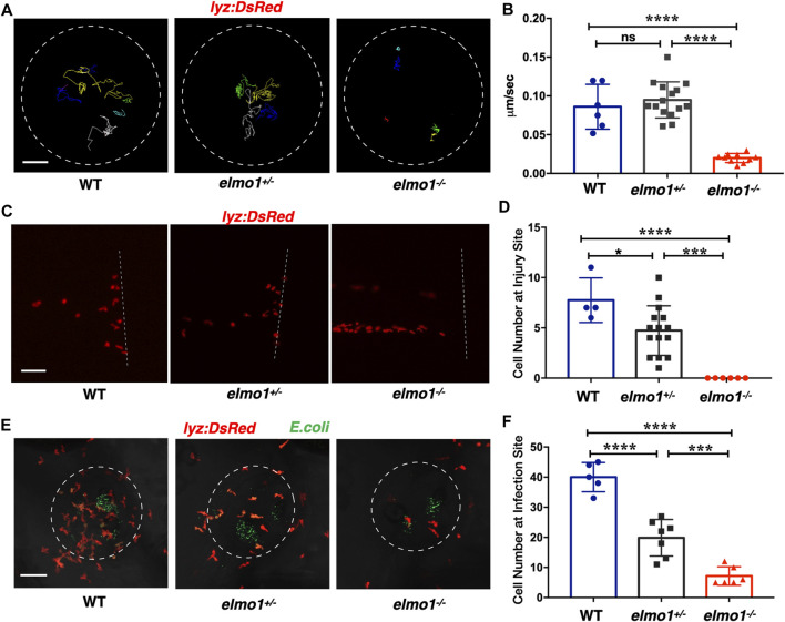
FIGURE 3 Neutrophils showed attenuated motility and impaired chemotaxis to injury/infection in the elmo1 mutant. (A) Track path of lyz:DsRed labeled neutrophils of the WT, elmo1+/? and elmo1?/? larvae on the yolk sac recorded by live imaging at 3 dpf. The white dotted circle indicates the imaging region of the yolk sac. Each line represents the migration path of individual cells. Scale bar: 50 ?m. (B) Quantification of neutrophils migration speed of the WT (6 cells of 3 larvae), elmo1+/? (15 cells of 5 larvae), and elmo1?/? (10 cells of 5 larvae). The elmo1?/? showed dramatically decreased speed compared with the WT and elmo1+/?. (C) Fluorescent image of tail fin transection of the WT, elmo1+/? and elmo1?/? larvae at 3 dpf. The larvae tail region was imaged and the white dotted line represents the transection site. lyz:DsRed represents neutrophils failed to accumulate to the injury site in the elmo1?/? compared with the WT and elmo1+/?. Scale bar: 50 ?m. (D) Quantification of neutrophils accumulated at the tail fin transection site. Neutrophils of the elmo1?/? larvae failed to respond to injury compared with the WT and elmo1+/?. (E) Fluorescent image of bacterial infection in the otic vesicle of WT, elmo1+/? and elmo1?/? larvae at 3 dpf. The white dotted circle represents the infection region. Bacteria of E.coli were labeled by GFP. lyz:DsRed labeled neutrophils showed a decreasing number at the infection region in the elmo1?/? compared with the WT and elmo1+/?. Scale bar: 50 ?m. (F) Quantification of lyz:DsRed labeled neutrophils accumulated at the infection region. Neutrophils of the elmo1?/? larvae failed to respond to infection compared with the WT and elmo1+/?. In quantification results, each dot represents the neutrophil number in the infected region in individual larvae. Three independent experiments were performed. Here presents one result of three experiments. One-way ANOVA, ns: no significance, *p < 0.05, ***p < 0.005, ****p < 0.001. (B, C, F).
Image
Figure Caption
Figure Data
Acknowledgments
This image is the copyrighted work of the attributed author or publisher, and
ZFIN has permission only to display this image to its users.
Additional permissions should be obtained from the applicable author or publisher of the image.
Full text @ Front Cell Dev Biol

