
|
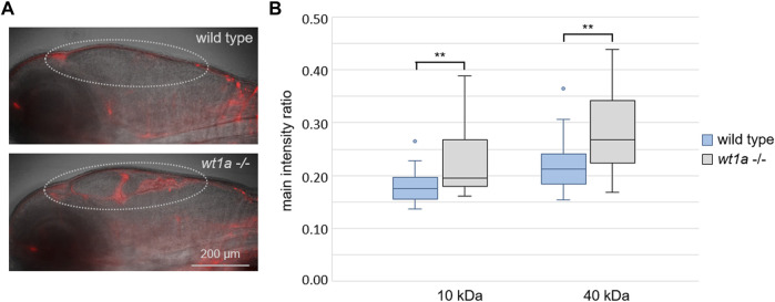
FIGURE 5
Wt1a inactivation reduces the blood-CSF barrier function. Fluorescently labeled dextrans were injected into the cardinal vein of 5 days old larvae of the wt1a ex1_del5 line and leakage into the ventricles was analyzed. (A) Representative overlay images of transmission and rhodamin-dextran (10 KDa) fluorescence recorded by light sheet microscopy of larvae that were fixed 4 h after injection. The dotted line marks the ventricular system. (B) Ratios of fluorescence intensities of rhodamin-dextran (10 KDa) and fluorescein-dextran (40 kDa) in the ventriclular region relative to the respective fluorescence in the pupil were estimated in vivo 40 min post injections. n = 24 for wild types and 22 for mutants; p-values according to ANOVA: 0.003 and 0.002 (**) for 10 and 40 kDa, respectively.