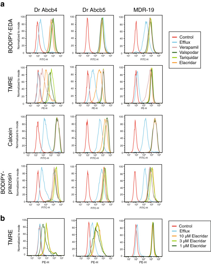
Figure 3 Zebrafish Abcb4 and Abcb5 differentially transport fluorescent P-gp substrates. (a) HEK293 cells transfected to express zebrafish Abcb4 (Dr Abcb4), zebrafish Abcb5 (Dr Abcb5) or human P-gp (MDR-19) were incubated in medium with 0.5 然 BODIPY FL-EDA, 0.5 然 TMRE, 150 nM calcein-AM or 0.5 然 BODIPY prazosin in the presence or absence of 10 然 elacridar, 10 然 tariquidar, 10 然 valspodar, or 100 然 verapamil for 30 min. The medium was removed and replaced with substrate-free medium in the presence or absence of inhibitor for an additional 1 h. Cells were incubated with fluorescent substrate alone, yielding the Efflux histogram and cell autofluorescence is depicted by the Control histogram. (b) Cells from (a) were incubated with medium containing 0.5 然 TMRE in the presence or absence of 1, 3, or 10 然 elacridar for 30 min after which the medium was removed and replaced with substrate-free medium in the presence or absence of inhibitor for an additional 1 h.
Image
Figure Caption
Acknowledgments
This image is the copyrighted work of the attributed author or publisher, and
ZFIN has permission only to display this image to its users.
Additional permissions should be obtained from the applicable author or publisher of the image.
Full text @ Sci. Rep.

