
|
Fig. 6.
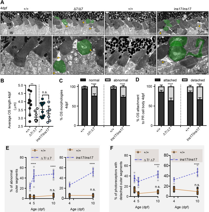
Photoreceptors in pcdh15b mutants develop with defects that progressively worsen with age. (A) Representative TEM images of the central ONL (top row) and OS (bottom row) in wild-type siblings, ?7 and ins17 mutants at 4 dpf. Green represents deformed/abnormal OSs. Yellow arrowheads point to regions of disconnection between the IS and OS. (B-D) Quantification of the average length of all OSs in the retina per section (B), the proportion of OS morphologies (C) and the percentage of attached/detached OSs (D) in wild-type siblings, ?7 and ins17 mutants at 4 dpf. The entire ONL was used for quantification. (E,F) Developmental trend of the percentage of abnormal OSs (E) and photoreceptors with detached OSs (F) in the retina from 4 dpf to 10 dpf in wild-type siblings, ?7 or ins17 mutants. Statistical analyses were performed using Student's t-tests (unpaired, two-tailed) (B-D) and two-way ANOVA (E,F). n.s., not significant; **P?0.01, ****P?0.0001. Number of eyes/OSs analysed for 4 dpf data: +/+, 10 eyes/1528 OSs; ?7/?7, 8 eyes/1412 OSs; +/+, 10 eyes/1829 OSs; ins17/ins17, 6 eyes/1328 OSs. IS, inner segment; ONL, outer nuclear layer; OS, outer segment.