
|
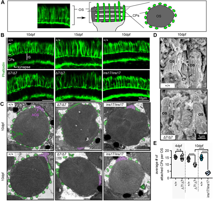
Fig. 3.
CPs are formed in pcdh15b mutants but are weakly attached and reduced/lost over time. (A) Schematic illustrating CPs surrounding the OS of photoreceptors. The F-actin stain, phalloidin, is used to label CPs. Transverse sections along the length of the photoreceptor show long CPs along their length, whereas horizontal cross-sections through the OS show OS surrounded by a ?ring? of CPs. In all images, CPs are coloured green. (B) Representative images of phalloidin staining in the photoreceptor layer in wild-type siblings (+/+) and ?7 or ins17 mutants at 10 dpf and 15 dpf (n=8+ individuals for each genotype). (C) Representative TEM images of horizontal cross-sections through the OS in wild-type siblings and ?7 and ins17 mutants. CPs (connected or disconnected to the OS) are coloured green; AOSs are coloured magenta. (D) Representative SEM images show CPs along the OS in wild-type siblings, which are absent/decreased in ?7 mutants (5 eyes from n=5 individuals per genotype). (E) Quantification of the average number of connected CPs per OS in wild-type siblings and pcdh15b mutants at 4 dpf and 10 dpf. OSs quantified were sampled from the central retina in the ONL. Number of eyes analysed for CP counts: 4 dpf: +/+, 5 eyes; ?7/?7, 8 eyes; 10 dpf: +/+, 6 eyes; ?7/?7, 6 eyes; +/+, 8 eyes; ins17/ins17, 8 eyes. Statistical analyses in E were performed using Student's t-test (unpaired, two-tailed) for 4 dpf and one-way ANOVA for 10 dpf. n.s., not significant; ****P?0.0001. AOS, accessory outer segment; CP, calyceal process; IS, inner segment; OS, outer segment.