Image
Figure Caption
Fig 6
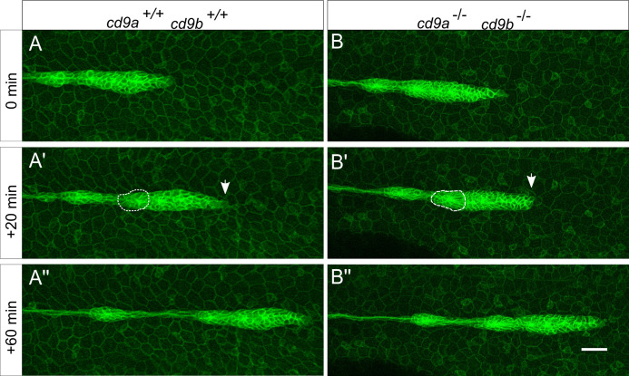
Primordium organisation appears normal in cd9 dKO(cldnb:gfp) embryos from 30 hpf.
A-B: Still images from a time-lapse recording of a migrating primordium in (a) WT and (b) cd9 dKOs. 0 minutes shows initial deposition as a proneuromast becomes distinct from the primordium and then 2 sequential images show (a?-b?) 20 minutes and (a??-b??) 60 minutes later. In the primordium of both WT and cd9 dKOs, filopodia can be seen at the leading edge (white arrow) and the formation of rosettes in the trailing edge (white dashed circle). Representative images from two videos that included two depositions each. Scale bar: 20 ?m.
Acknowledgments
This image is the copyrighted work of the attributed author or publisher, and
ZFIN has permission only to display this image to its users.
Additional permissions should be obtained from the applicable author or publisher of the image.
Full text @ PLoS One

