Figure 5
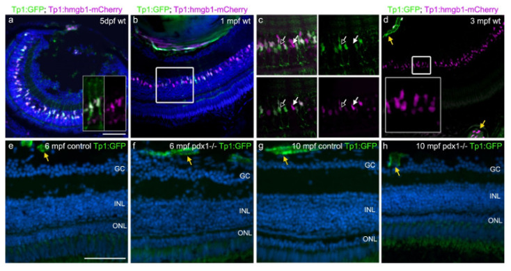
Notch signaling is not re-activated during chronic hyperglycemia. (a?d) Tg(tp1:GFP;tp1:hmgb1-mCherry) double transgenic 5 dpf larvae (a) and adults (b?d) showed gradual loss of GFP expression in the INL over time, while hmgb1-mCherry expression labeled cells in the INL through adulthood. In some GFP-positive cells at 5 dpf, projections extend along the apical?basal axis (a, inset). In (c), which shows a close-up of the boxed region in (b), GFP and mCherry variably overlap in INL nuclei. At 3 mpf, INL cells show variable levels of mCherry, and are GFP negative (d, inset). GFP and mCherry are coexpressed in retinal and choroidal vessels (d, yellow arrows). (e?h) In 6 mpf (e,f) and 10 mpf (g,h) control and pdx1?/? mutants, GFP expression via notch-responsive elements cannot be detected in the neural retina. Expression is limited to retinal and choroidal vessels (yellow arrows). Sections were counterstained with DAPI (blue). Scale bar: 50 Ám. (GC, ganglion cell layer; INL, inner nuclear layer; ONL, outer nuclear layer).

