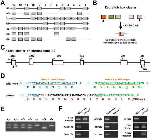
Fig. 1 Zebrafishhoxclusters and isolation of hoxaa cluster-deleted mutants. (A) The zebrafish possesses 48 hox genes in seven hox clusters. (B) Strategy for deletion of the genomic region corresponding to the hox cluster. Two dgRNAs (arrowheads), which recognize hox genes located at both ends of hox clusters, were used to delete the target genomic region. (C) Schematic of the hoxaa cluster locus. Six hox genes in the hoxaa cluster are depicted as white rectangles. The arrow shows the orientation of the transcription of each hox gene. Two black arrowheads indicate the genomic loci of the target sequence of dgRNAs, which were used to delete the genomic region of the hoxaa cluster. (D) Large genomic deletion in the hoxaa cluster mutant. The flanking sequences of two dgRNAs-target are shown. The target sequence of hoxaa-5? dgRNA is exon 1 of hoxa13a (blue letters), and hoxaa-3? dgRNA is targeted to exon 1 of hoxa1a (green letters). The hoxaa mutant possessing a ?56.4 kb deletion and a 27 bp insertion (black letters) was isolated. This indel mutation resulted in frameshift of hoxa13a. (E) PCR-based genotyping of embryos obtained by intercross between hoxaa+/? fish. Genotyping was carried out with the three primers shown by blue arrowheads in C. The sequences of primers and PCR cycles are shown in Table S3. (F) Specific deletion of the hoxaa cluster was confirmed by PCR. The genomic DNA extracted from wild-type and hoxaa?/? embryos was used as a template for PCR. The sequences of the primers used in this analysis are listed in Table S4.
Image
Figure Caption
Acknowledgments
This image is the copyrighted work of the attributed author or publisher, and
ZFIN has permission only to display this image to its users.
Additional permissions should be obtained from the applicable author or publisher of the image.
Full text @ Development

