
|
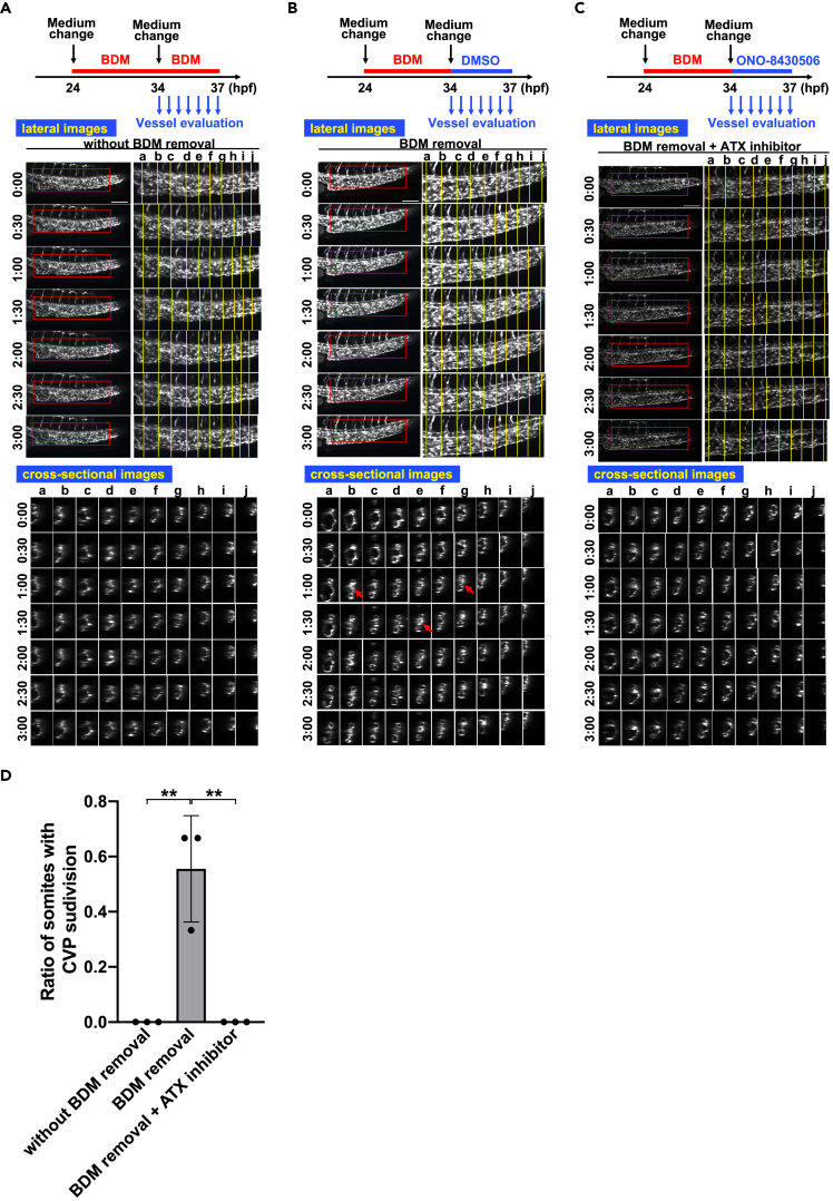
Figure 7
Blood flow-induced CVP formation is dependent on ATX
(A) Twenty-four hpf embryos were pre-treated with BDM (12 mM) for ten hours by changing the medium to a medium containing BDM, and at 34 hpf the medium was changed to the same medium containing BDM and the sequential time-lapse images were taken for 3 h every 30 min. The CVP structures did not change significantly for 3 h. Scale bars, 100 μm.
(B) Twenty-four hpf embryos were pre-treated with BDM as in (A), and at 34 hpf the medium was changed to a medium without BDM and the sequential time-lapse images were taken for 3 h every 30 min. Note that subdivisions of CVP accompanied by constriction of vessels were observed (arrows). Scale bars, 100 μm.
(C) Twenty-four hpf embryos were pre-treated with BDM as in (A), and at 34 hpf the medium was changed to a medium without BDM but containing ONO-8430506, and the sequential time-lapse images were taken for 3 h every 30 min. Note that subdivisions and constriction of CVP were significantly suppressed. Scale bars, 100 μm.
(D) Ratio of somites with CVP subdivision was quantified by evaluating ten somites (somite a–j in
See also