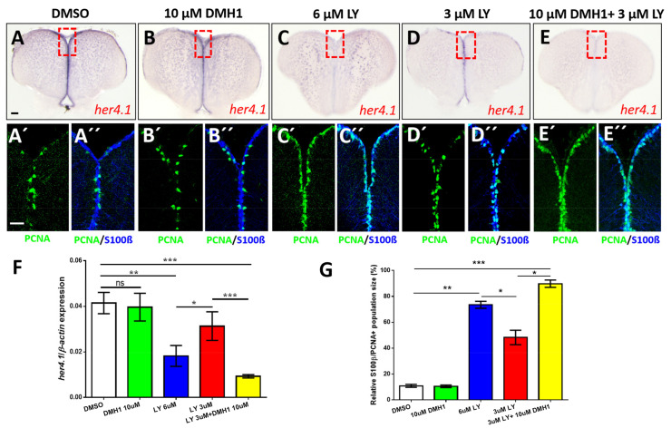
Figure 7 Notch and BMP signaling pathways interact to control quiescence of NSCs. (A?E) Expression of her4.1 revealed by ISH on control telencephala (DMSO, A) or telencephala treated with different concentrations of DMH1 (B), LY411575 (LY, C,D), or a combination of LY and DMH1 (E). (B) 10 ?M DMH1 does not influence strongly the expression of her4.1. (C) Notch blockage reduces her4.1 expression along the ventricular zone. (D) After reduction of the concentration of LY to 3 ?M, her4.1 expression is still recognizable in the ventricular zone. (E) Combination of 3 ?M LY with 10 ?M DMH1 blocks her4.1 expression in the ventricular zone suggesting that the two pathways interact. Red rectangles (A?E) illustrate regions of immunostaining in A??E?. (A??E?) Cross-sections of the pallial ventricular zone following different concentrations of DMH1 or LY treatment or a combination of both, immunostained for the NSC marker S100? (blue) and the proliferation marker PCNA (green). The proportion of PCNA+/S100?+ cells is increased in the groups treated with 6 ?M LY alone (C?,C??) or treated with a combination of 3 ?M LY and 10 ?M DMH1 (E?,E??). (F) RT-qPCR analysis of her4.1 mRNA expression under different conditions of drug treatment. (G) Quantification of the relative population size of PCNA+/S100?+ cells under different conditions. Significance is indicated by asterisks: ns, not significant; * 0.01 ? p < 0.05; ** p < 0.01; *** p < 0.001. Scale bars: 20 ?m (A?,A??,B?,B??,C?,C??,D?,D??,E?,E??), 100 ?m (A,B,C,D,E).
Image
Figure Caption
Figure Data
Acknowledgments
This image is the copyrighted work of the attributed author or publisher, and
ZFIN has permission only to display this image to its users.
Additional permissions should be obtained from the applicable author or publisher of the image.
Full text @ Cells

