IMAGE

Fig. 3

ID
ZDB-IMAGE-211025-178
Genes
Source
Figures for Ribera et al., 2021
Image
Figure Caption

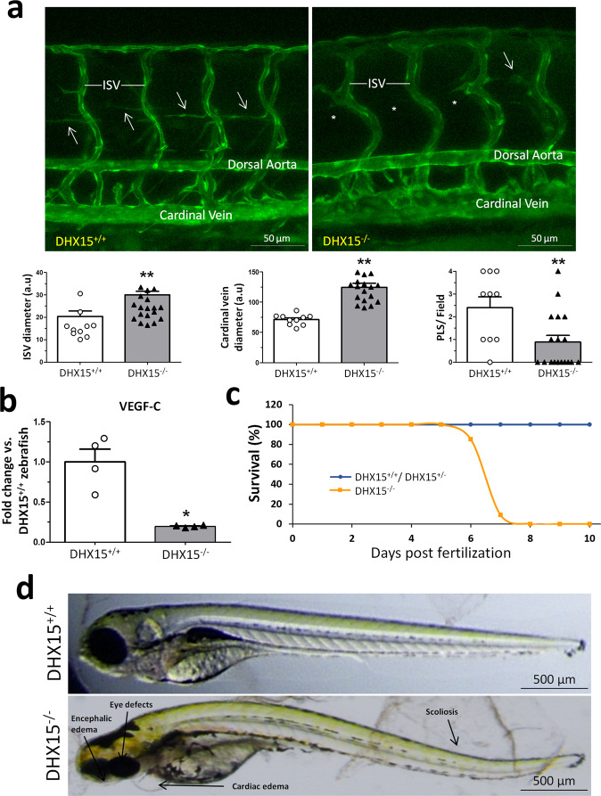
Fig. 3 Characterization of embryonic vascular anomalies associated with <italic>DHX15</italic> gene deficient in zebrafish.

a Representative vascular images of DHX15+/+ and DHX15−/− larvae at 5 day post fertilization (dpf) revealing a reduced formation of the parachordal line (arrows). Asterisks denote the absence of these vascular structures in DHX15−/− animals. Quantifications of cardinal vein diameter, intersegmental vessels (ISV), and number of parachordal lymphangioblast strings (PLS) are shown in the graphs. **p < 0.01 vs. wild-type zebrafish, unpaired two-tailed Student’s t-test (n = 10 and n = 18 for the DHX15+/+ and DHX15−/− conditions, respectively). Arbitrary units: a.u. b RNA extraction of zebrafish embryos at 4 dpf from either wild-type or DHX15 knockout larvae was performed. mRNA expression was analyzed by RT-qPCR. The graph shows the different expression levels of the VEGF-C gene in the DHX15+/+ and DHX15−/− conditions. mRNA levels are shown as fold change relative to HPRT mRNA levels. *p < 0.05 vs. wild-type unpaired two-tailed Student’s t-test (n = 4 biologically independent samples for each condition). c Survival assessment assay. The graph shows the larvae survival rate through the first 10 dpf according to their different genotype (n = 15 zebrafish larvae for each condition). d Representative images comparing wild-type and DHX15−/− larvae at 7 dpf where morphological defects including encephalic and cardiac edema, scoliosis, and impaired neural/eye growth are evident. All bar graphs are presented as mean ± SEM.

Figure Data
Acknowledgments
This image is the copyrighted work of the attributed author or publisher, and ZFIN has permission only to display this image to its users. Additional permissions should be obtained from the applicable author or publisher of the image. Full text @ Commun Biol