
|
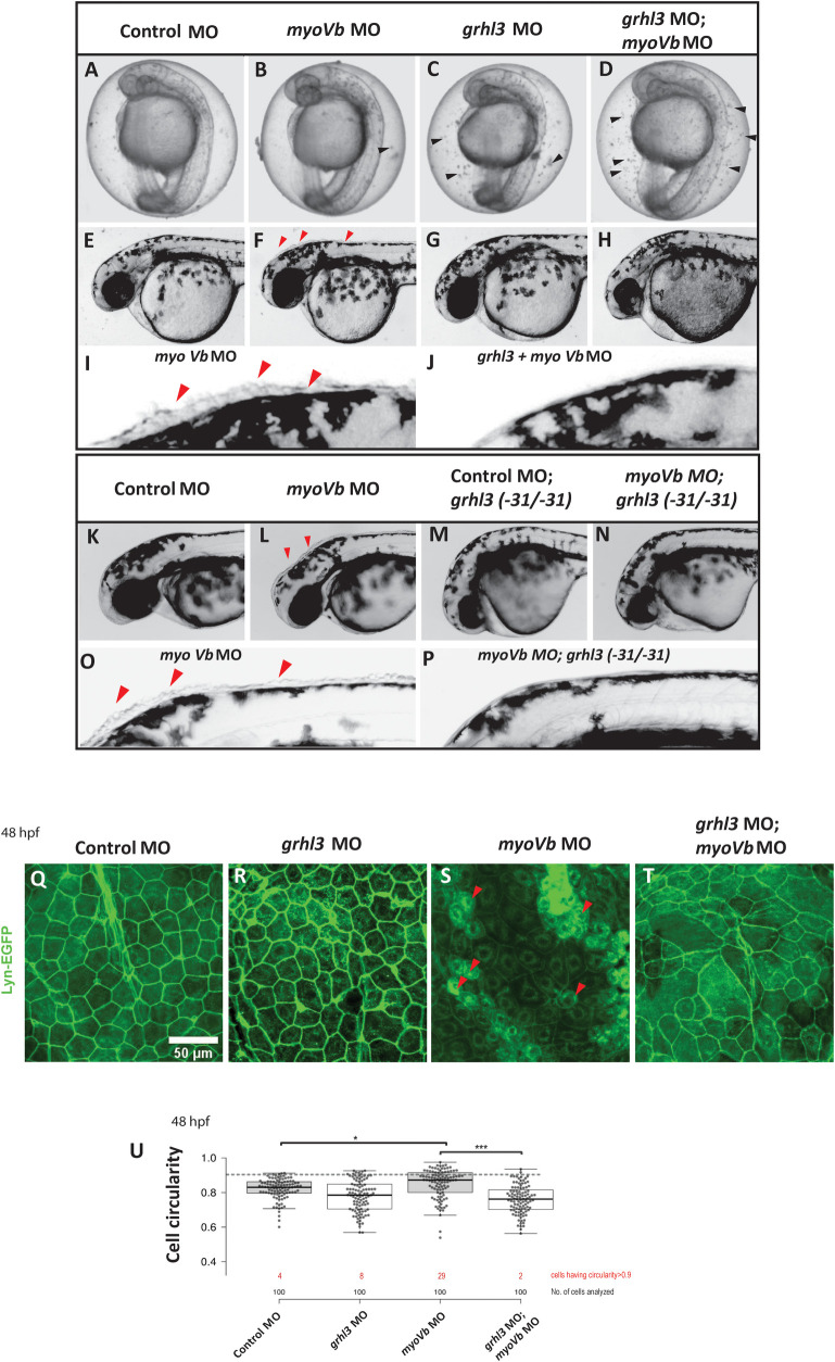
Fig 4
Bright field images of embryos at 28 hpf injected with control MO (A), myoVb MO (B), grhl3 MO (C), and grhl3 MO; myoVb MO (D). Black arrowheads indicate cell debris accumulated inside the chorion of grhl3 and grhl3;myoVb double morphant embryos, presumably due to epidermal cell shedding. Representative images of the head region of zebrafish larvae injected with control MO (E), myoVb MO (F), grhl3 MO (G), and grhl3;myoVb double morphant (H) at 36 hpf. Zoomed in images of the dorsal head region of myoVb morphant (I) and grhl3;myoVb double morphant (J) embryos, respectively. Representative images of wild-type siblings injected with control MO (K) and myoVb MO (L) and grhl3(-31/-31) mutants injected with control MO (M) and myoVb MO (N). Zoomed in images of dorsal head region of wild-type siblings (O) and grhl3 (-31/-31) mutants (P) injected with myoVb MO. Note the presence of rounded cells in myoVb MO in F, I, L, O?indicated by red arrowheads?and their absence in myoVb; grhl3 double deficient embryos in H, J, N, P. Representative confocal images of the peridermal cells from the stated genetic conditions at 48hpf (Q-T). Note the presence of rounded cells in myoVb MO indicated by red triangles. Box and whisker plot (U) showing quantitation of cell circularity in control, grhl3 MO, myoVb MO, and grhl3;myoVb MO at 48hpf. Each column represents cell circularities of 25 cells each from four embryos per condition (total 100). Grey dotted line represents the cell circularity threshold used for classifying round cells. Red numbers indicate % cells having cell circularity values above threshold. myoVb MO embryos show significantly higher median cell circularity than that in control and grhl3;myoVb MO (Kruskal Wallis One way ANOVA p<0.05). For all comparisons and statistical significance please refer to S2 Data.