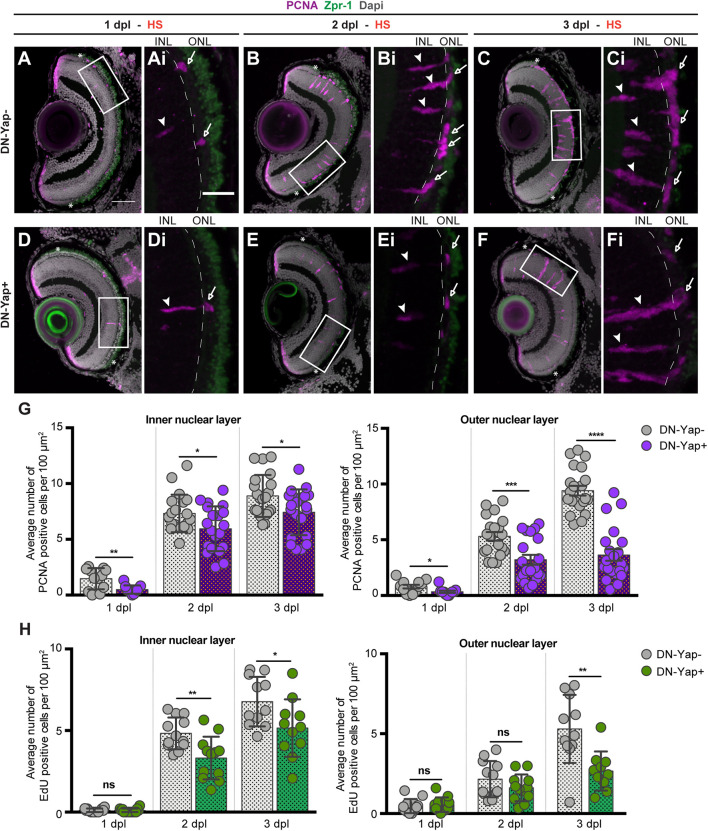
Yap inhibition reduces cell proliferation in the retina after photoreceptor-induced light lesion. (A–F) Transverse cryosections of DN-Yap− (A–C) and DN-Yap+ (D–F) retinas immunostained for proliferating cell nuclear antigen (PCNA) (magenta) and Zpr-1 (green) and counterstained with DAPI (gray). White arrowheads and arrows indicate PCNA+ cells in the inner nuclear layer (INL) and outer nuclear layer (ONL), respectively, at 1 dpl (A,D magnified, Ai,Di), 2 dpl (B,E, magnified, Bi,Ei), and 3 dpl (C,F, magnified, Ci,Fi). (G) Quantification of PCNA + cells in DN-Yap− and DN-Yap + larva retinas from 1 to 3 dpl, in the INL and ONL. (H) Quantification of EdU + cells in DN-Yap− and DN-Yap + larva retinas from 1 to 3 dpl, in the INL and ONL. ns, non-significant; *p < 0.05, **p < 0.01, ***p < 0.001, ****p < 0.0001. Unpaired t-test with Welch’s correction. Asterisks delimitate the lesioned region. Dashed lines delimitate INL from ONL. White boxes delimitate magnified (Ai–Fi). Scale bars correspond to 50 μm in (A,C), and 20 μm in magnified (Ai,Ci).
Acknowledgments
This image is the copyrighted work of the attributed author or publisher, and
ZFIN has permission only to display this image to its users.
Additional permissions should be obtained from the applicable author or publisher of the image.
Full text @ Front Cell Dev Biol
Your Input Welcome
Thank you for submitting comments. Your input has been emailed to ZFIN curators who may contact you if
additional information is required.
Oops. Something went wrong. Please try again later.