IMAGE

Fig 2

ID
ZDB-IMAGE-210926-17
Antibodies
Source
Figures for Suzzi et al., 2021
Image
Figure Caption

Fig 2 Early but transient neurodevelopmental phenotypes in mzLrrk2 larvae.

(A and B) Increased cell death in the brains of mzLrrk2 larvae (5 dpf) as revealed by TUNEL assay. Quantification was carried out over the whole brain, subdivided into anterior (telencephalon), middle (diencephalon, mesencephalon), and posterior (rhombencephalon) portions. (A) Scale bar: 100 μm. (B) Plot represents means ± s.e.m. Statistical analyses: two-tailed Student’s t-test. (C-E) No evidence of midbrain neurodegeneration in mzLrrk2 larvae (5 dpf). HuC/D IHC (C-top and D) and acetylated (Ac.) Tubulin IHC (C-bottom and E) were used to label postmitotic neurons and axonal network. (C) Scale bar: 100 μm. (E) IR, immunoreactivity (measured as integrated density). (D and E) Plots represent means ± s.e.m. Statistical analyses: two-tailed Student’s t-test. (F-H) Claudin k+ myelinated commissural fibers in the ventral hindbrain were reduced in mzLrrk2 larvae (5 dpf; F and G), but not in youngsters (1 mo; H; representative images in S6 Fig). (F) Scale bar: 100 μm. (H) IR, immunoreactivity (measured as integrated density). (G and H) Plots represent means ± s.e.m. Statistical analyses: two-tailed Student’s t-test. (I-K) L-Plastin+ microglia/leukocytes were reduced in the brains of mzLrrk2 larvae (5 dpf; I and J), but not in the brains of adult fish (6 mo; K, quantification in the telencephalon as representative brain region; representative images in S8 Fig). (I) Scale bar: 100 μm. (J) Quantification of microglia/leukocytes in the larval brain was performed after image segmentation as depicted in S7A–S7D Fig. (J and K) Plots represent means ± s.e.m. Statistical analyses: two-tailed Student’s t-test.
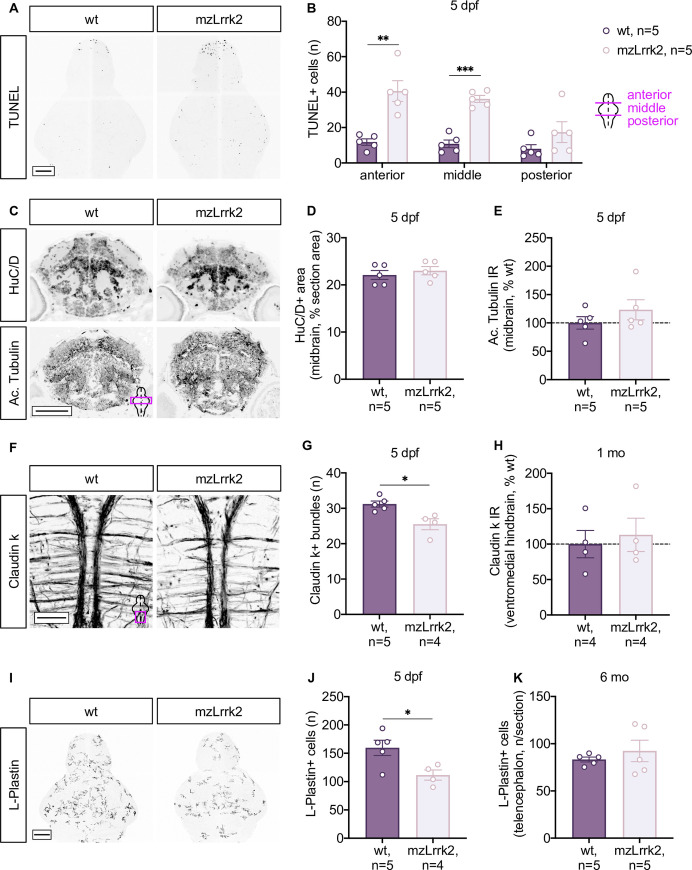
Figure Data
Acknowledgments
This image is the copyrighted work of the attributed author or publisher, and ZFIN has permission only to display this image to its users. Additional permissions should be obtained from the applicable author or publisher of the image. Full text @ PLoS Genet.