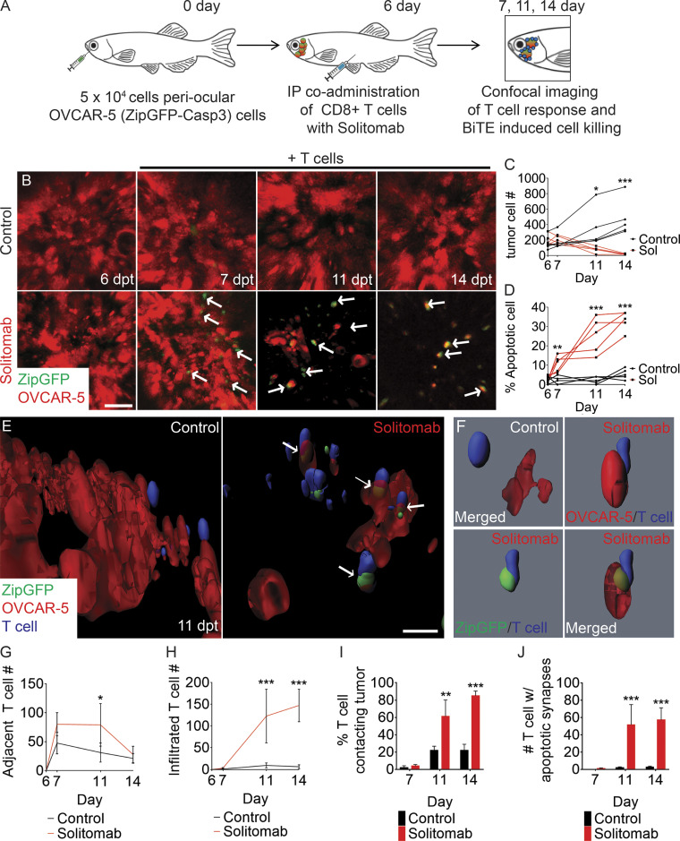
Figure 4.
Single-cell quantitation of apoptotic immune synapse formation following BiTE immunotherapy in ovarian carcinoma. (A) Schematic of experimental design. (B) Serial imaging of animals engrafted with OVCAR-5 cells engineered to express mCherry and ZipGFP-Casp3 before (6 dpt) and after IP injection of CFSE-labeled CD8+ T cells administered with either EpCAM antibody (control) or EpCAM/CD3 solitomab (7, 11, and 14 dpt; arrows denote apoptotic cells). (C) Quantification of tumor cell numbers in engrafted animals over time. (D) Quantification of apoptotic cells over time. (E) 3D modeling showing control EpCAM antibody (left) or CD3/EpCAM solitomab (right) imaged at 11 dpt (white arrows denote apoptotic cells engaged with T cells). (F) Single-cell renderings showing immune synapse formation in real time. Control (top left panel) and experiment (top right and bottom panels). (G and H) T cell migration to sites adjacent to the tumor (G) and infiltrated into the tumor mass (H; 0.1 mm3 volume). (I) Quantification of the percentage of T cells contacting tumor cells. (J) Number of tumor-infiltrating T cells that contain apoptotic synapses with tumor cells. 0.0125 mm3 volume. *, P < 0.05; **, P < 0.01; ***, P < 0.001, Student?s t test. Scale bar equals 10 µm (B and E). n = 5 fish/experimental arm for all analyses shown. Error bars denote ±SD. Sol, solitomab.

