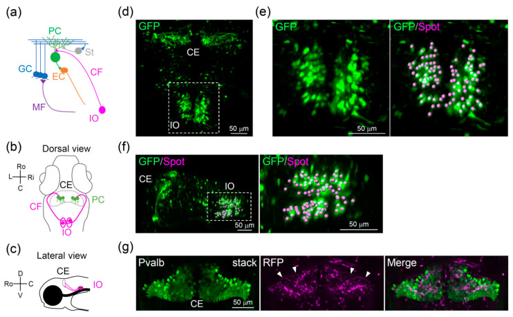
Figure 1
Cerebellar circuits in zebrafish: (a) Schematic diagram of the cerebellar circuits in zebrafish. (b,c) Schematic diagram of the olivocerebellar circuits: (b) Dorsal view and (c) lateral view. (d?f) Distribution of inferior olive neurons in Tg(hspGFFDMC28C;UAS:GFP) zebrafish at 7 days post-fertilization (dpf): (d,e) Dorsal view, with a high-magnification image shown in (e), where spots indicate the position of the soma of inferior olive neurons. (f) Dorsolateral view, with a high-magnification image of the inferior olive shown in the right panel. (g) Dorsal view of the cerebellum of Tg(hspGFFDMC28C;UAS:RFP) larva stained with Parvalbumin 7 (Pvalb) at 6 dpf (confocal z-stack images). Arrowheads indicate CFs. CE, cerebellum; CF, climbing fiber; EC, eurydendroid cell; GC, granule cell; IO, inferior olive; MF, mossy fiber; PC, Purkinje cell; St, stellate cell; Ro, rostral; C, cordal; L, left; Ri, right; D, dorsal; V, ventral.

