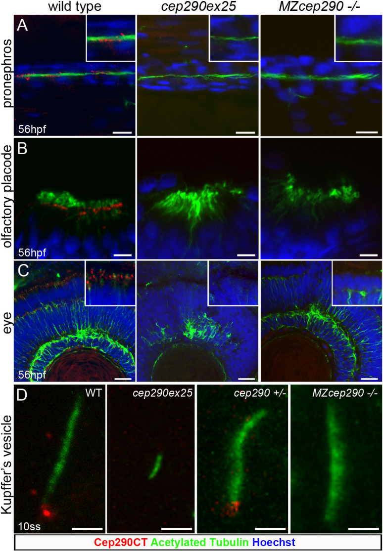
IMAGE

Fig. 2.

ID
ZDB-IMAGE-210814-56
Genes
Antibodies
Source
Figures for Cardenas-Rodriguez et al., 2021
Image
Figure Caption

Fig. 2.

Functional loss of Cep290 in zebrafish morphants and mutant embryos. (A-C) Cep290CT antibody staining was lost in pronephric tubule multicilia bundles (A), at the base of nose multicilia (B) and in photoreceptor, outer and inner plexiform retina layers (C) upon injection of the cep290ex25 morpholino or in MZcep290−/− mutants. Loss of Cep290 expression caused an alteration of retina lamination. No effect was observed on pronephros and olfactory placode cilia structure. (D) Kupffer's vesicle cilia were significantly shorter in 10ss cep290ex25 morphant embryos compared to stage-matched embryos injected with control morpholino. By contrast, 10ss MZcep290 mutant embryos did not show shorter Kupffer's vesicle cilia compared with heterozygous sibling embryos. Images are representative of >8 embryos/biological replicates. Experiments were replicated a minimum of three times. Scale bars: 10 µm (A); 5 µm (B); 20 µm (C); 2 µm (D).

Figure Data
Acknowledgments
This image is the copyrighted work of the attributed author or publisher, and ZFIN has permission only to display this image to its users. Additional permissions should be obtained from the applicable author or publisher of the image. Full text @ J. Cell Sci.