
Image
|
Figure Caption
Fig 1
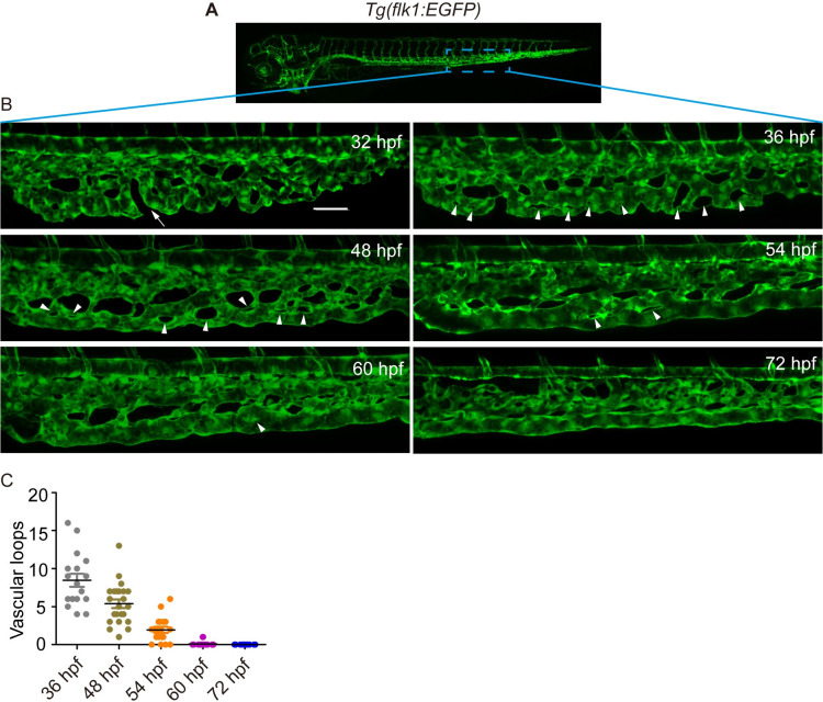
(A) A sketch map of the region. (B) Confocal images of the caudal vasculature of zebrafish embryos. The arrow points to ventral CVP capillaries without connection. Arrowheads point to vascular loops. Scale bar: 50 μm. (C) Quantification of the number of vascular loops in the CV: 36 hpf, n = 17 embryos; 48 hpf, n = 23 embryos; 54 hpf, n = 19 embryos; 60 hpf, n = 22 embryos; 72 hpf, n = 13 embryos.
Figure Data
Acknowledgments
This image is the copyrighted work of the attributed author or publisher, and
ZFIN has permission only to display this image to its users.
Additional permissions should be obtained from the applicable author or publisher of the image.
Full text @ PLoS Genet.