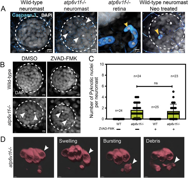
Fig. 6. (A) Cleaved Caspase 3 staining was absent in wild-type and atp6v1f?/? mutant neuromasts at 4?dpf, but was detected in the retina of atp6v1f?/? embryos, and in wild-type hair cells treated with neomycin (Neo) that induces Caspase 3 activation (yellow arrowheads). Nuclei were detected using DAPI. White arrowheads indicate pyknotic nuclei. (B) DAPI staining of neuromast nuclei at 4?dpf in wild-type and atp6v1f?/? embryos treated with either DMSO (control) or 300?ÁM ZVAD-FMK from 2?dpf to 4?dpf. White arrowheads indicate pyknotic nuclei. Dashed line circles indicate the neuromast boundary. Scale bars: 5 Ám. (C) Quantification of pyknotic nuclei per neuromast in control and 300?ÁM ZVAD-FMK-treated embryos. n=number of embryos. ns, not significant by unpaired Student's t-test with Welch's correction. (D) 3D rendering of time-lapse snapshots of Tg(myo6b:tdtomato); atp6v1f?/? hair cells undergoing necrosis-like morphological changes. The white arrowhead follows one hair cell swelling and then bursting over time. Scale bars: 3 Ám.
Image
Figure Caption
Figure Data
Acknowledgments
This image is the copyrighted work of the attributed author or publisher, and
ZFIN has permission only to display this image to its users.
Additional permissions should be obtained from the applicable author or publisher of the image.
Full text @ Dis. Model. Mech.

