IMAGE

Fig. 3.

ID
ZDB-IMAGE-210801-89
Genes
Antibodies
Source
Figures for Santra et al., 2021
Image
Figure Caption

Fig. 3.

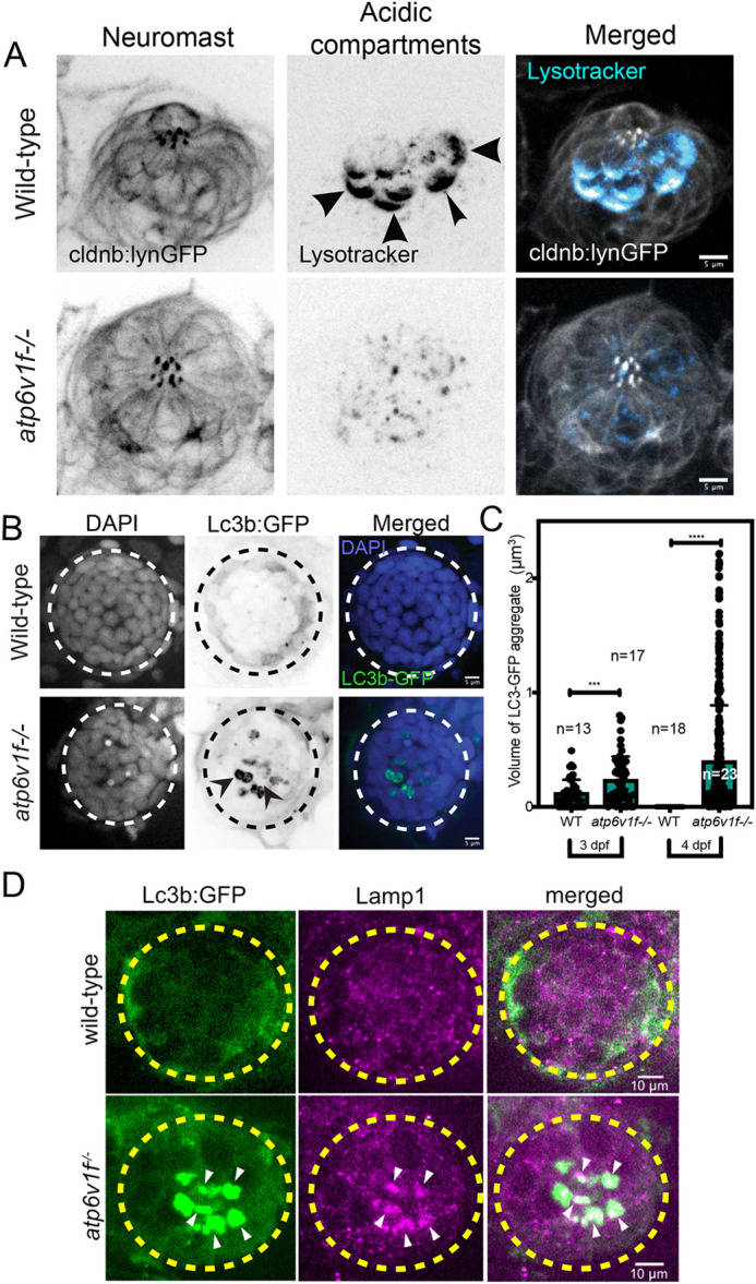
V-ATPase loss of function alters pH and induces autophagy defects in neuromasts. (A) Lysotracker staining in a wild-type neuromast at 4 dpf was reduced in atp6v1f−/− mutants. Neuromast cells are marked by Tg(cldnb:lynEGFP) transgene expression. Arrowheads point out basal accumulation of Lysotracker in wild-type hair cells. Scale bars: 5 µm. (B) Wild-type and atp6v1f−/− mutant neuromasts at 4 dpf stained for Lc3b-GFP (green) and nuclei using DAPI (blue). Arrowheads point out Lc3b-GFP aggregates. White and black dashed line circles indicate the neuromast boundary. (C) Quantification of volume of individual Lc3b-GFP aggregates in neuromasts from wild-type and atp6v1f−/− embryos. The number of data points reflects the finding that each embryo can have multiple neuromast aggregates. n=number of embryos. ***P=0.0005 and ****P<0.0001 by unpaired Student's t-test with Welch's correction. (D) Anti-Lamp1 antibody staining shows colocalization with Lc3b-GFP aggregates in atp6v1f−/− mutant neuromast. Yellow dashed line circles indicate hair cell cluster within a neuromast.

Figure Data
Acknowledgments
This image is the copyrighted work of the attributed author or publisher, and ZFIN has permission only to display this image to its users. Additional permissions should be obtained from the applicable author or publisher of the image. Full text @ Dis. Model. Mech.