IMAGE

Figure 9—figure supplement 2.

ID
ZDB-IMAGE-210725-99
Source
Figures for Ma et al., 2021
Image
Figure Caption

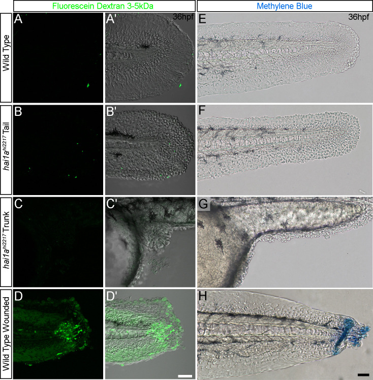
Figure 9—figure supplement 2. The barrier function of the <italic toggle='yes'>hai1a</italic> epidermis is not grossly compromised.

(A–D′) Lateral confocal images of the tails (AB′; D, D′) and trunk (C, C′) of 36hpf WT (AA′; DD′) and hai1ahi2217 embryos (B–C′) immersed in 2.5 mg/ml of 3–5 kDa fluorescein dextran for 30 min before brief destaining. Fluorescent images (A–D) are also shown overlaying DIC images (A′–D′). (E, F) Lateral DIC images of the tails (E, F, H) and trunk (G) of 36hpf WT (E, H) and hai1ahi2217 embryos (F, G) immersed in 0.075% of methylene blue for 30 min before brief destaining. Tails of WT embryos in (H) were wounded by scalpel cut before immersion. Scale bars: (D′, H) = 50 µm.

Acknowledgments
This image is the copyrighted work of the attributed author or publisher, and ZFIN has permission only to display this image to its users. Additional permissions should be obtained from the applicable author or publisher of the image. Full text @ Elife