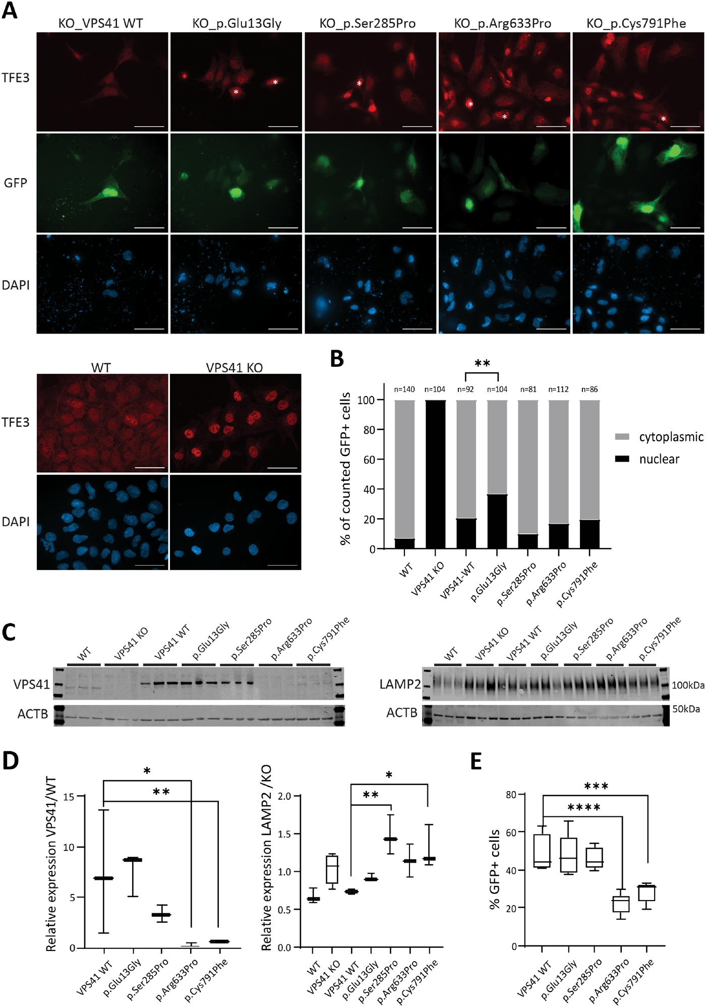
Fig. 2 VPS41 variants contribute to impaired lysosomal functions in vitro.(A) Immunocytochemistry assessing TFE3 in rescue experiments using transient expression of wild-type (WT) and mutant VPS41-t2a-GFP in VPS41 KO ESC. Transfected cells are marked by GFP and GFP+ cells showing no TFE3 relocalization to the cytoplasm are marked with white asterisks. Nuclei are counterstained with DAPI. Scale bars = 50 µm. (B) Quantification of A counting a minimum of 80 GFP+ cells from three replicates. ***P = 0.0001, binomial test (expected distribution based on relocalization upon transfection with the wild-type construct). (C) Western blotting detecting VPS41 (endogenous VPS41 99 kDa, V5-tag-VPS41 105 kDa, input 30 µg) and LAMP2 (100–120 kDa, input 20 µg), in wild-type and VPS41 KO ESCs and VPS41 KO ESCs transiently rescued with wild-type or mutant VPS41. (D) Quantification of C, *P < 0.05; **P < 0.01; ***P < 0.001 one-way ANOVA, multiple comparison test of mutant constructs to the wild-type. (E) Percentage of GFP+ cells upon transient transfections of VPS41-KO cells with VPS41-GFP plasmid spiked with mCherry, expressing wild-type or mutant VPS41. ***P = 0.0006, ****P < 0.0001, one-way ANOVA, multiple comparison test of mutant constructs to the wild-type; >17 000 cells were analysed per sample.
Image
Figure Caption
Acknowledgments
This image is the copyrighted work of the attributed author or publisher, and
ZFIN has permission only to display this image to its users.
Additional permissions should be obtained from the applicable author or publisher of the image.
Full text @ Brain

