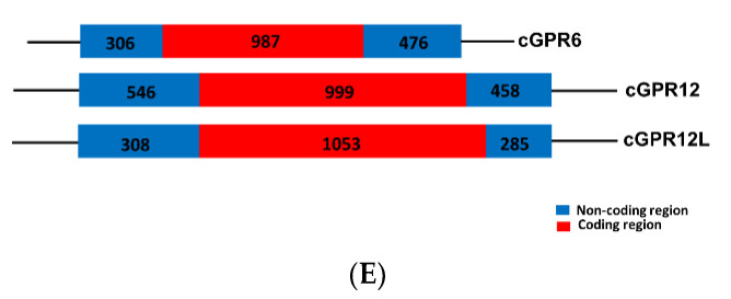
Figure 1 Amino acid sequence alignment of G protein-coupled receptor 3 (GPR3), GPR6, GPR12, and GPR12L in vertebrates. (A) Alignment of the cloned duck (d-)/zebrafish (zf-)/pig (p-) GPR3 with that of humans (hGPR3, NP_005272.1), giant pandas (gpGPR3, XP_034503372.1) and spotted gars (sgGPR3, XP_015204111.1); (B) Alignment of the cloned chicken(c-)/duck(d-)/zebrafish(zf-)/pig(p-) GPR6 with that of humans (hGPR6, NP_005275.1), giant pandas (gpGPR6, XP_002925693.2), and spotted gars (sgGPR6, XP_006626355.1); (C) Alignment of the cloned chicken (c-)/duck (d-)/zebrafish (zf-)/pig (p-) GPR12 with that of humans (hGPR12, NP_005279.1), giant pandas (gpGPR12, XP_002924260.1), and spotted gars (sgGPR12, XP_015196669.1); (D) Alignment of the cloned chicken (c-)/duck (d-)/zebrafish (zf-) GPR12L with that of anole lizard (lGPR12L, XP_028571577.1) and spotted gars (sgGPR12L, XP_015206591.1). The seven transmembrane domains (TMD1?7) and N-glycosylation sites (NXT/S) are shaded; the XXXWD motif is boxed; conserved motifs (DRY, XWXP, and NPXXY) are marked by arrowheads; (E) Exon?intron organization of chicken GPR6, GPR12, and GPR12L. The red boxes indicate the coding region, and the numbers inside denote the size (in bp); while the blue box represents the untranslated regions, (UTRs) and the number within specifies the size (in bp).
Image
Figure Caption
Acknowledgments
This image is the copyrighted work of the attributed author or publisher, and
ZFIN has permission only to display this image to its users.
Additional permissions should be obtained from the applicable author or publisher of the image.
Full text @ Genes (Basel)

