
|
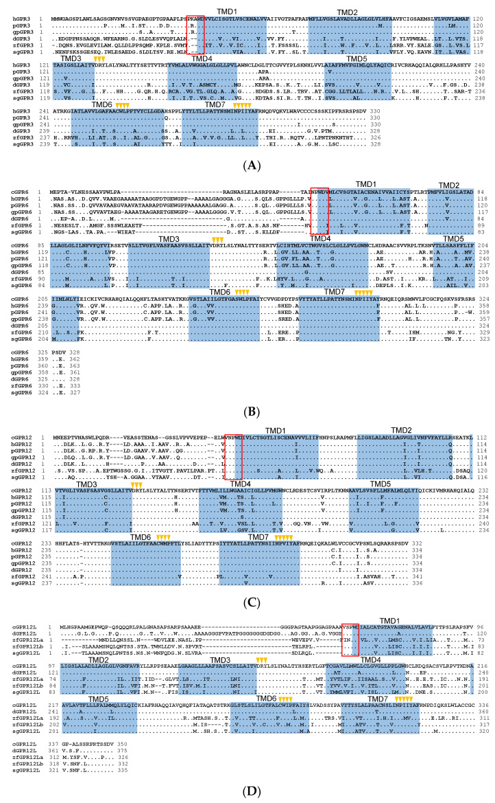
Figure 1 Amino acid sequence alignment of G protein-coupled receptor 3 (GPR3), GPR6, GPR12, and GPR12L in vertebrates. (A) Alignment of the cloned duck (d-)/zebrafish (zf-)/pig (p-) GPR3 with that of humans (hGPR3, NP_005272.1), giant pandas (gpGPR3, XP_034503372.1) and spotted gars (sgGPR3, XP_015204111.1); (B) Alignment of the cloned chicken(c-)/duck(d-)/zebrafish(zf-)/pig(p-) GPR6 with that of humans (hGPR6, NP_005275.1), giant pandas (gpGPR6, XP_002925693.2), and spotted gars (sgGPR6, XP_006626355.1); (C) Alignment of the cloned chicken (c-)/duck (d-)/zebrafish (zf-)/pig (p-) GPR12 with that of humans (hGPR12, NP_005279.1), giant pandas (gpGPR12, XP_002924260.1), and spotted gars (sgGPR12, XP_015196669.1); (D) Alignment of the cloned chicken (c-)/duck (d-)/zebrafish (zf-) GPR12L with that of anole lizard (lGPR12L, XP_028571577.1) and spotted gars (sgGPR12L, XP_015206591.1). The seven transmembrane domains (TMD1?7) and N-glycosylation sites (NXT/S) are shaded; the XXXWD motif is boxed; conserved motifs (DRY, XWXP, and NPXXY) are marked by arrowheads; (E) Exon?intron organization of chicken GPR6, GPR12, and GPR12L. The red boxes indicate the coding region, and the numbers inside denote the size (in bp); while the blue box represents the untranslated regions, (UTRs) and the number within specifies the size (in bp).