
Image
|
Figure Caption
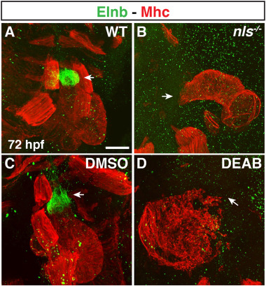
Fig. 7 Smooth muscle within the OFT is absent in RA-deficient embryos. (A?D) IHC of Elnb (green) and Mhc (red) in hearts of (A,B) WT sibling and nls mutant embryos and (C,D) DMSO- and DEAB-treated embryos at 72 hpf. Number of embryos examined per condition: WT n ?= ?6, nls n ?= ?8, DMSO-treated n ?= ?12, DEAB-treated n ?= ?9. Views are lateral with arterial pole of the heart up. Arrows indicate the Elnb in WT sibling and DMSO-treated and the lack of Elnb in nls mutant and DEAB-treated at the arterial poles of the hearts. Scale bar ? 50 ??m.
Acknowledgments
This image is the copyrighted work of the attributed author or publisher, and
ZFIN has permission only to display this image to its users.
Additional permissions should be obtained from the applicable author or publisher of the image.
Reprinted from Developmental Biology, 473, Duong, T.B., Holowiecki, A., Waxman, J.S., Retinoic acid signaling restricts the size of the first heart field within the anterior lateral plate mesoderm, 119-129, Copyright (2021) with permission from Elsevier. Full text @ Dev. Biol.