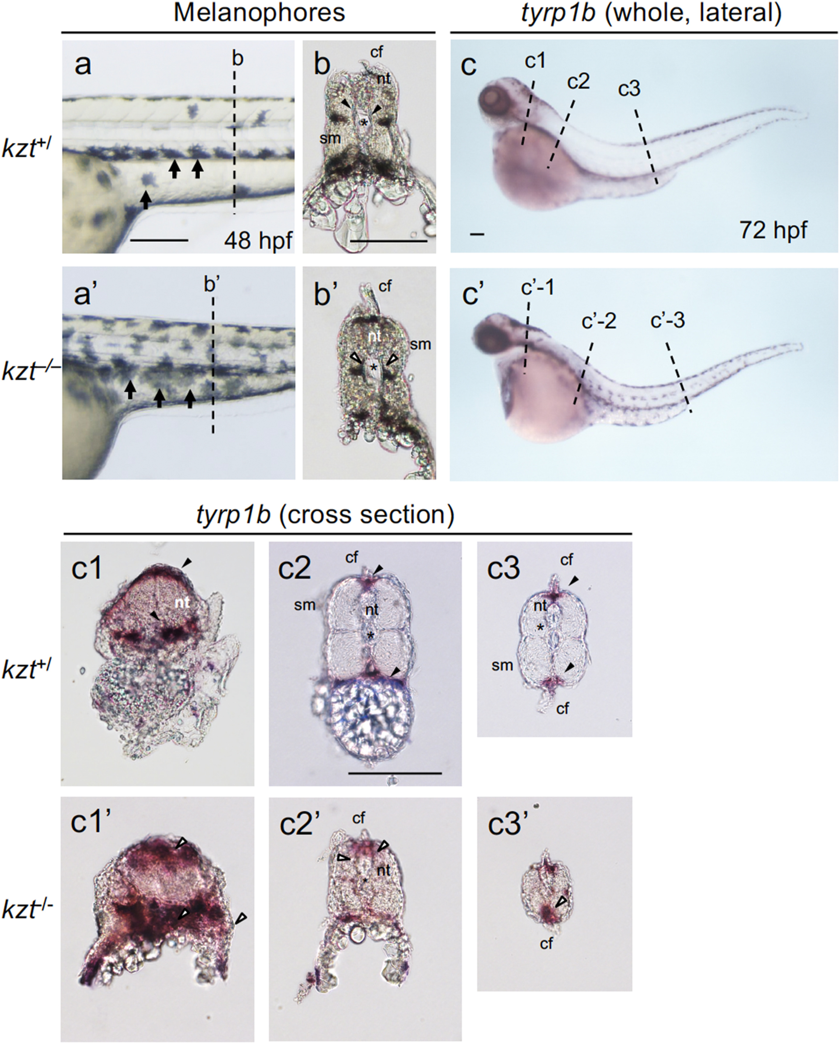
Fig. 5 Migration and pattern formation of melanophores are abnormal in kzt embryos. (a, b, a?, b?) Distribution of melanophores in sibling (kzt+/) and homozygous (kzt?/?) embryos. (a, a?) Lateral views of the trunks with anterior to the left and dorsal to the top, showing the distribution of melanophores (small arrows) at 48 hpf. (b?, b?) Cross-sections of fixed embryos at positions corresponding to the broken lines in (a) and (a?), respectively, showing melanophore distribution within embryonic bodies. In wild-type embryos, melanophores were absent in the vicinity (solid arrowheads) of the notochord (asterisk), whereas they were distributed on the surface of the notochord in kzt mutants (open arrowheads). (c, c?) The expression of tyrp1b localized by whole-mount in situ hybridization (WISH), identifying melanophores in sibling embryos (c, kzt+/?) and homozygotes (c?, kzt?/?). (c1?c3, c1??c3?) Cross-sections of stained embryos at positions corresponding to the broken lines in (c) and (c?), respectively, showing melanophore distribution (arrowheads). In wild-type embryos, melanophore distribution was restricted beneath the epidermis and in the dorsal-most and ventral-most regions (solid arrowheads), whereas melanophores were abnormally distributed on the surface of the notochord and within embryos in a dorsoventrally broad manner in kzt mutants (open arrowheads). cf, caudal fin; nt, neural tube; sm, somite; ?, notochord. Scale bars, 200 ??m.
Reprinted from Developmental Biology, 472, Takahashi, K., Ito, Y., Yoshimura, M., Nikaido, M., Yuikawa, T., Kawamura, A., Tsuda, S., Kage, D., Yamasu, K., A globin-family protein, cytoglobin 1, is involved in the development of neural crest-derived tissues and organs in zebrafish, 1-17, Copyright (2020) with permission from Elsevier. Full text @ Dev. Biol.

