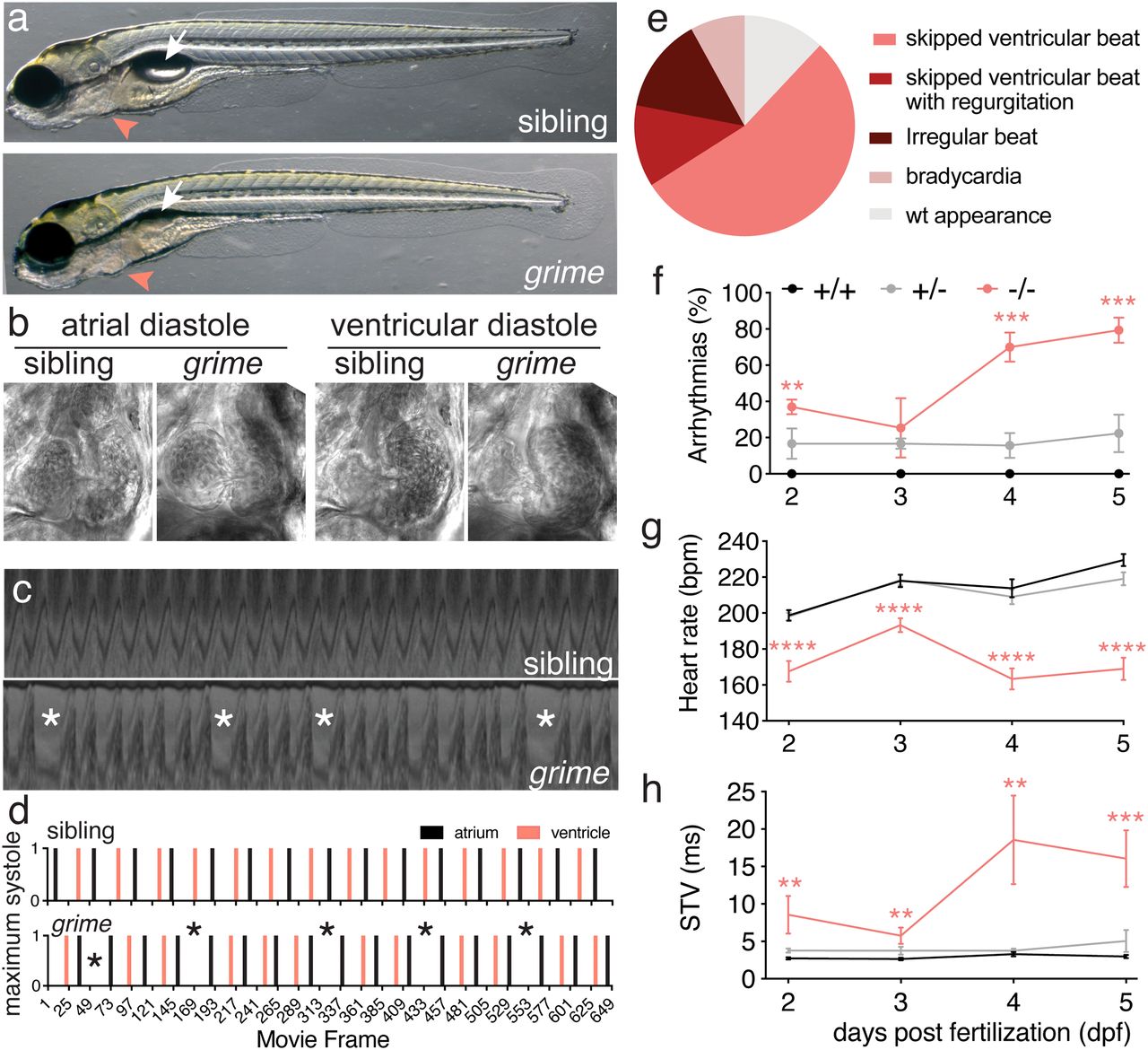
Fig. 1 grime mutants display specific cardiac arrhythmia. (A) Brightfield lateral view images of wild-type and grime (tmem161buq4ks) mutant zebrafish at 5 dpf. The swim bladder of grime mutants often fails to inflate (white arrow) but embryos are otherwise indistinguishable (location of the heart depicted by pink arrowhead). (B) Frontal view images of live heart by brightfield, high-speed imaging showing sibling and grime mutant hearts are anatomically similar both at atrial and ventricular diastole. (C) Kymograph of atrial movement versus time from high-speed movies of sibling and grime heartbeat. Mutant example shows an embryo with irregular heart rate (single asterisks). (D) Graph depicting maximum systole of atrium (black) and ventricle (pink) as a function of time in sibling and grime mutants taken from examples Movies S1 and S2. The mutant example shows the skipped ventricular beat phenotype (single asterisks). (E) Summary of phenotypes observed in the grime mutants at 5 dpf. (F?H) Graphs of wild type, heterozygous, and homozygous grime mutants at 2 to 5 dpf, showing (F) the percentage of embryos presenting with arrhythmias (for more detail, see SI Appendix, Table S1) (G) heart rates, and (H) the STV in atrial contraction interval (mean ± SEM; n = 20 to 47; **P ? 0.01, ***P ? 0.001, ****P ? 0.0001).
Image
Figure Caption
Figure Data
Acknowledgments
This image is the copyrighted work of the attributed author or publisher, and
ZFIN has permission only to display this image to its users.
Additional permissions should be obtained from the applicable author or publisher of the image.
Full text @ Proc. Natl. Acad. Sci. USA

