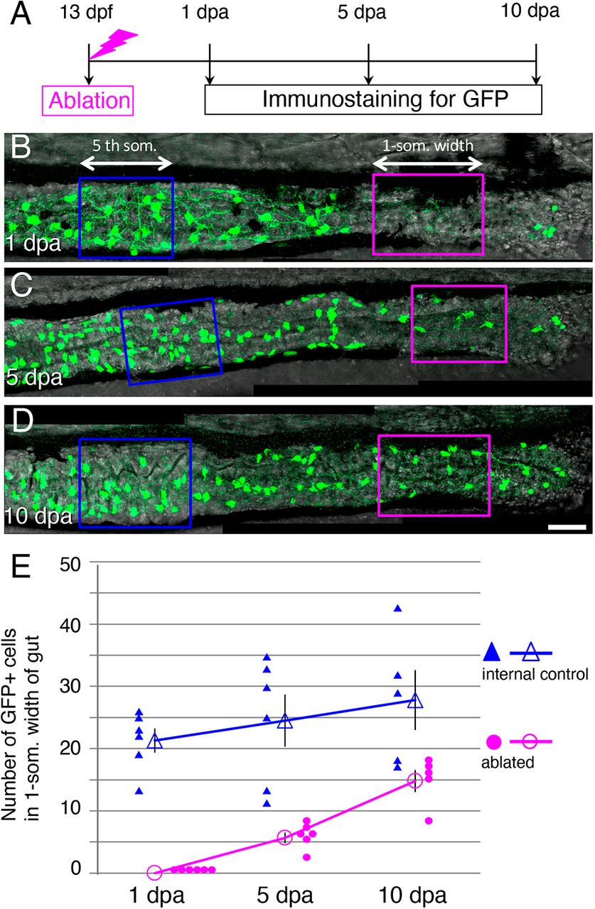
Fig. 3 The number of GFP+ cells in the ablated area increases after ablation. (A) Timeline of the experiments. After ablation at 13?dpf, larvae were cultured and fixed at 1, 5 and 10?dpa for immunostaining. (B-D) Posterior intestines stained for GFP at 1?dpa (B) 5 dpa (C), and 10?dpa (D). The arrows and enclosed areas indicate the one-somite-width of intestine used for counting the GFP+ cells in the ablated area (magenta; ablated) and unablated (blue; internal control) area of the same larva at the second- and the fifth-somite level, respectively (see Materials and Methods for details). Images are left side views of the mid-distal intestines, with the anterior end positioned to the left of the image. Scale bar: 50?µm. (E) Number of GFP+ cells in ablated and internal control areas at 1, 5 and 10?dpa. Filled symbols indicate the numbers in individual larvae, and outline symbols indicate the meanħs.e.m. In the ablated areas, the number of GFP+ cells increased and reached ?50% of the control level within 10?days, whereas a significant change in the number of cells was not observed in the control area during the same period (1?dpa versus 10?dpa; two-tailed unpaired t-test, P=0.2).
Image
Figure Caption
Acknowledgments
This image is the copyrighted work of the attributed author or publisher, and
ZFIN has permission only to display this image to its users.
Additional permissions should be obtained from the applicable author or publisher of the image.
Full text @ Development

