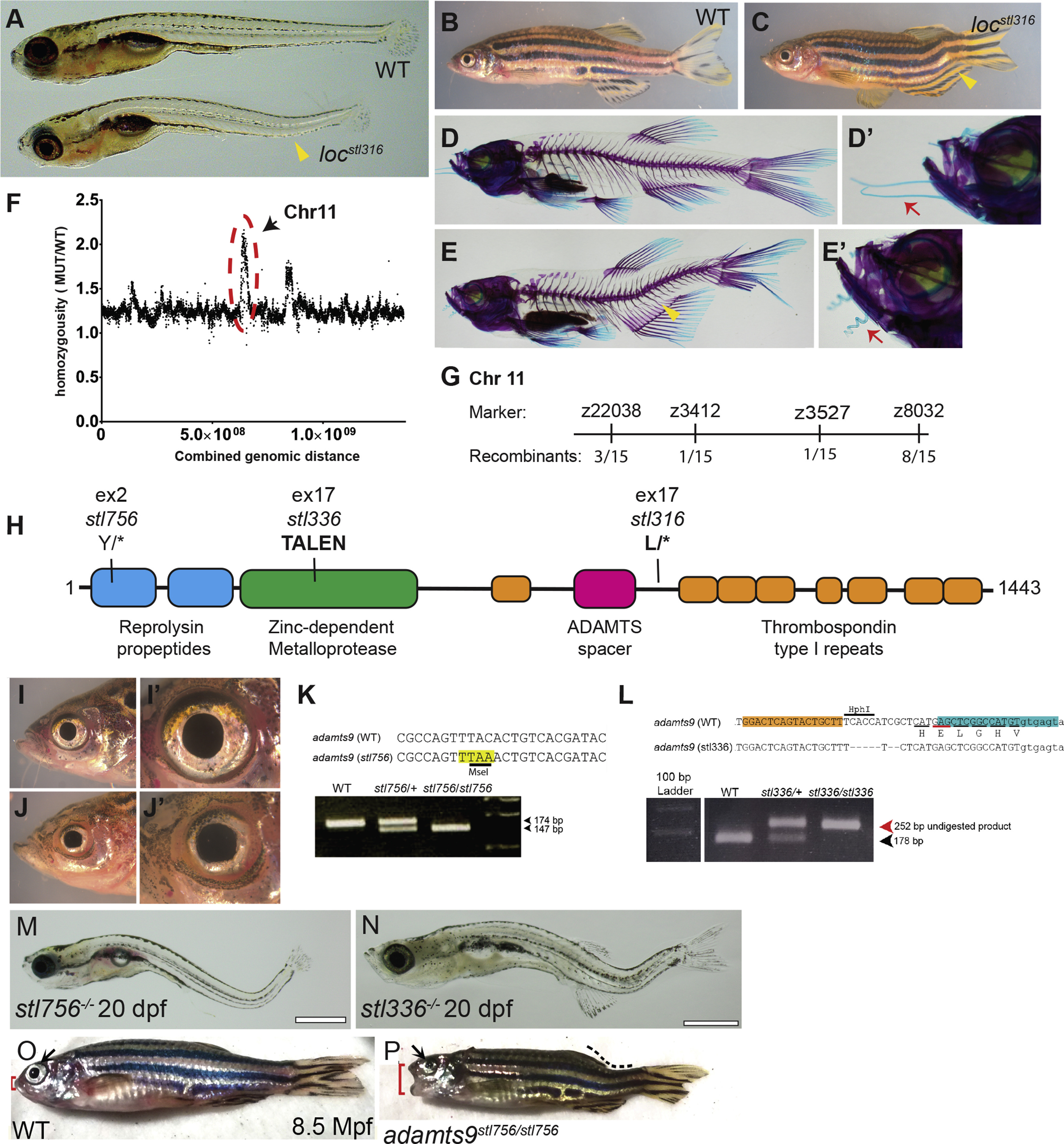
Fig. 8 The pimienta locura/locstl97 mutation is predicted to generate a truncated Adamts9 protein. Bright field of representative sibling WT and mutant locstl316/stl316 phenotypes at 5 dpf (A) and 60 dpf (B, C) and Alizarin-Red and Alcian-Blue skeletal preparations of WT (D, D?) and locstl316/stl316 mutants (E, E?) highlighting the caudal-localized curvature of the notochord and spine (yellow arrowhead). Close-ups of zebrafish faces highlighting phenotypic differences in barbel morphology (red arrows) normally straight in the wild type (D?) and curly or kinky in locstl316/stl316 mutants (E?). Homozygosity based mapping graphed as mutant allele ratio over the total genomic distance shows a major peak of homozygosity at Chromosome 11 and a minor peak at Chromosome 15 (F). The locstl316 lesion was mapped to a region of Chromosome 11 between markers z3412 and z3527 (number of recombinants are denoted at each marker) representing a large ~26 ?Mb region annotated for 519 genes in the Zv10 build. A schematic of the predicted 1443-amino acid protein, showing the locations of three novel adamts9 mutant alleles in this study (H). (I-J?) Close-ups brightfield imaging of head and eye to highlight the alteration in eye morphology observed in a representative adamts9stl316/stl316 mutant (J, J?) and wild-type (I, I?). (M?P) Bright field imaging shows severe curvatures of the spine and eye defects are observed by 20 dpf for both (M) adamts9stl756 and (N) adamts9stl336. (P) While most of these mutants are larval lethal rare escapers can survive to adulthood but display severe defects in eye development (black arrow), spine curvature (dotted line), and defects of the jaw (red bracket), (O) not observed in WT siblings.
Reprinted from Developmental Biology, 471, Gray, R.S., Gonzalez, R., Ackerman, S.D., Minowa, R., Griest, J.F., Bayrak, M.N., Troutwine, B., Canter, S., Monk, K.R., Sepich, D.S., Solnica-Krezel, L., Postembryonic screen for mutations affecting spine development in zebrafish, 18-33, Copyright (2020) with permission from Elsevier. Full text @ Dev. Biol.

