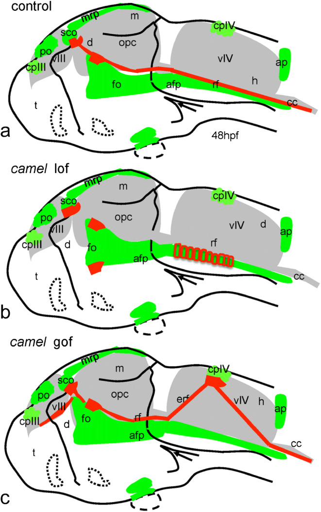
Schematics show organization of the Reissner fiber in respect of the ventricular system (based on Figs. 9, 10, and 11). a–c 48 hpf. a Controls. b Anti-Camel morpholino–mediated loss-of-function. c Camel mRNA–mediated gain-of-function. Green color, midline structures and some CVOs; red, RF and AFRU+ material. Abbreviations: afp, anterior floor plate; ap, area postrema; cc, central canal; cpIII, choroid plexus of the third ventricle; cpIV, choroid plexus of the fourth ventricle; d, diencephalon; e, epiphysis; erf, ectopic Reissner fiber; h, hindbrain; fo, flexural organ; fp, floor plate; m, midbrain; mrp, midbrain roof plate; opc, optocoele (Sylvius aqueduct); po, pineal organ; rf, Reissner fiber; sco, subcommissural organ; t, telencephalon; vIII, third ventricle; vIV, fourth ventricle
Acknowledgments
This image is the copyrighted work of the attributed author or publisher, and
ZFIN has permission only to display this image to its users.
Additional permissions should be obtained from the applicable author or publisher of the image.
Full text @ Cell Tissue Res.
Your Input Welcome
Thank you for submitting comments. Your input has been emailed to ZFIN curators who may contact you if
additional information is required.
Oops. Something went wrong. Please try again later.