IMAGE

Fig. 3.

ID
ZDB-IMAGE-210305-4
Source
Figures for Bastos de Oliveira et al., 2021
Image
Figure Caption

Fig. 3.

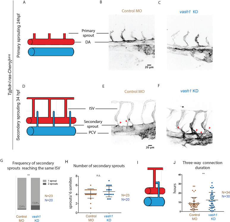
Vash1 deficient embryos exhibit aberrant secondary sprouting. (A-F) Primary and secondary sprouting in control and vash1 KD Tg[kdr-l:ras-Cherry]s916 zebrafish embryos, with labelled blood vessels. Primary sprouting occurs from the DA at 24-30 hpf (A-C). Secondary sprouting occurs from the PCV at 34-40 hpf and ISVs form (D-F). Red arrowheads indicate secondary sprouts (E,F). Pictures are representative of three replicated experiments. (G,H) Quantification of observed frequency that either one or two different sprouts reach an ISV (G) and of number of secondary sprouts per six somites (H). n=87 ISVs from 20 embryos for vash1 KD group, and n=82 ISVs from 23 control embryos, from three biological replicates. **P<0.0021 (Mann–Whitney test). n.s., non-significant. (I,J) A transient three-way connection is formed when the secondary sprout connects with the primary ISV and lumenizes (I), after which one of its branches is resolved. The duration of three-way connections from lumenization to resolution was quantified in control and vash1 MO-injected embryos (J). Data points represent 45 ISVs from 34 embryos for vash1 KD and 39 ISVs from 30 embryos for control groups, from four biological replicates. **P<0.0021 (Kolmogorov–Smirnov test). Data are median±s.d. (G) and mean±s.d. (H,J).

Figure Data
Acknowledgments
This image is the copyrighted work of the attributed author or publisher, and ZFIN has permission only to display this image to its users. Additional permissions should be obtained from the applicable author or publisher of the image. Full text @ Development