IMAGE

Fig. 1.

ID
ZDB-IMAGE-210305-2
Genes
Source
Figures for Bastos de Oliveira et al., 2021
Image
Figure Caption

Fig. 1.

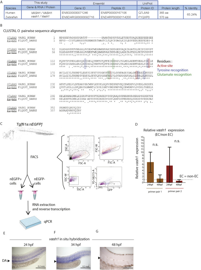
vash1 is highly expressed in zebrafish endothelium. (A,B) Amino acid sequence alignment using CLUSTAL O (Uniprot) of human and zebrafish Vash1 showing 65.24% sequence similarity (A), including in the indicated residues necessary for tubulin detyrosination (B). (C,D) Analysis of vash1 expression in zebrafish embryos at 24 and 48 hpf. FACS sorting and gating strategy for isolation of nEGFP+ and nEGFP− cells from Tg[fli1a:nEGFP]y7 embryos, their RNA extraction and reverse transcription leading to qPCR (C). vash1 expression, analysed by two independent primer pairs, shows a tendency towards being enriched in the endothelium of zebrafish embryos at 24 hpf (D). Data normalised to housekeeping genes. Data are mean±s.d. Each data point is an average of three technical replicates per sample, and each experiment has three biological replicates per developmental stage. n.s., not significantly different (Mann–Whitney test). (E-G) In situ hybridization of vash1 in the zebrafish embryo trunk at 24 hpf (E), 34 hpf (F) and 48 hpf (G). Arrowhead indicates where the dorsal aorta (DA) locates in the embryos. Pictures are representative of three replicated experiments. n=40 embryos for each developmental stage.

Figure Data
Acknowledgments
This image is the copyrighted work of the attributed author or publisher, and ZFIN has permission only to display this image to its users. Additional permissions should be obtained from the applicable author or publisher of the image. Full text @ Development