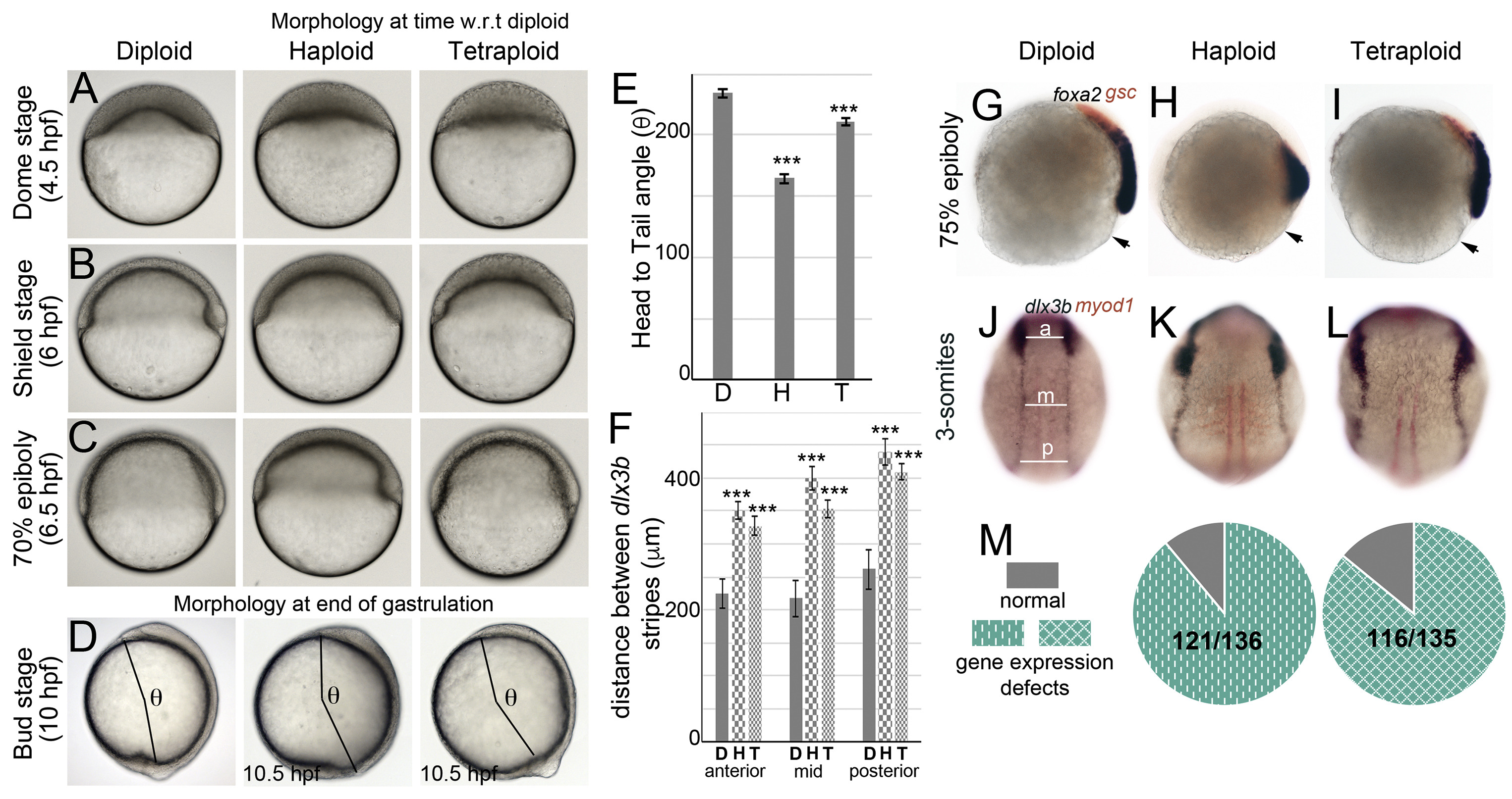
Fig. 3 Embryos with average cell sizes different than the norm manifest epiboly delay and dorsal convergence defects during gastrulation. Live DIC images during epiboly and gastrulation (A?D). Haploids and tetraploids initiate epiboly (A) and gastrulation (B) later than diploids and develop with a delay during gastrulation (C). Haploids and tetraploids conclude gastrulation with a delay and manifest a shorter body axis at bud stage, quantified as the head to tail angle (beginning of head to tailbud from the center of the yolk ball, denoted as ?, D). Quantification of ? in diploids (n ?= ?15), haploids (n ?= ?10), tetraploids (n ?= ?8) (E). RNA in situs for foxa2 (blue) and gsc (red) at 75% epiboly (G-I, diploid n ?= ?34/34, haploid n ?= ?62/63, tetraploid n ?= ?58/66, arrow indicates embryo margin). RNA in situs for dlx3b (blue) and myod1 (red) at 3-somites (J-L, diploid n ?= ?56/56, haploid n ?= ?63/77, tetraploid n ?= ?69/80). Quantification of the distance between dlx3b stripes along the AP axis at three sites anterior (a), mid (m) and posterior (p) as shown in J, for all conditions (F). Asterisks denote statistical significance based on unpaired t-test and One-way ANOVA (p ?? ?0.05), error bars represent Standard Error of Mean (SEM) for E and F. Cumulative quantification of all gastrulation defects (live and RNA in situ) in haploids and tetraploids (M).
Reprinted from Developmental Biology, 468(1-2), Menon, T., Borbora, A.S., Kumar, R., Nair, S., Dynamic optima in cell sizes during early development enable normal gastrulation in zebrafish embryos, 26-40, Copyright (2020) with permission from Elsevier. Full text @ Dev. Biol.

